Images and videos
Images


Epicondylitis
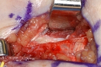
Surgery for refractory medial epicondylitis: pickups lifting off area of degenerative tendon after elliptical incision to excise this area
From the collection of Dr Brian Fitzgerald, Naval Medical Center San Diego, CA; used with permission
See this image in context in the following section/s:

Epicondylitis
AP radiograph of elbow with lateral calcification from chronic lateral epicondylitis
From the collection of Daniel J. Solomon, Naval Medical Center San Diego, CA; used with permission
See this image in context in the following section/s:

Epicondylitis
Markings for swing incision location for patient with chronic refractory medial epicondylitis
From the collection of Dr Brian Fitzgerald, Naval Medical Center San Diego, CA; used with permission
See this image in context in the following section/s:
Epicondylitis
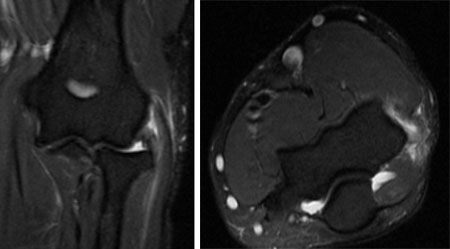
Coronal MRI and axial MRI in the same patient, showing high signal in extensor carpi radialis brevis
From the collection of Daniel J. Solomon, Naval Medical Center San Diego, CA; used with permission
See this image in context in the following section/s:

Epicondylitis
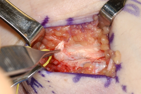
Surgery for refractory medial epicondylitis: probe placed on area of degenerative tendon showing loss of normal tendon appearance
From the collection of Dr Brian Fitzgerald, Naval Medical Center San Diego, CA; used with permission
See this image in context in the following section/s:

Epicondylitis
Surgery for refractory medial epicondylitis: medial epicondyle exposed
From the collection of Dr Brian Fitzgerald, Naval Medical Center San Diego, CA; used with permission
See this image in context in the following section/s:
Epicondylitis
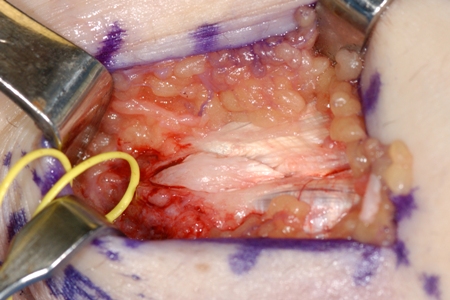
Surgery for refractory medial epicondylitis: degenerative tendon removed
From the collection of Dr Brian Fitzgerald, Naval Medical Center San Diego, CA; used with permission
See this image in context in the following section/s:
Use of this content is subject to our disclaimer