Images and videos
Images


Evaluation of vision loss
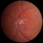
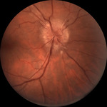
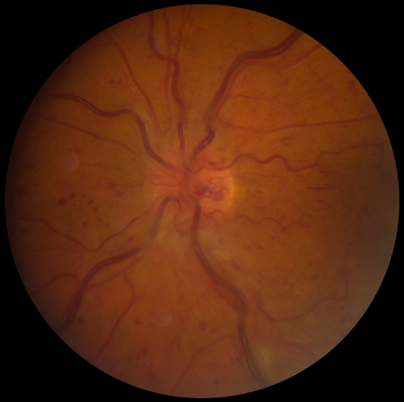
Segmental optic disk swelling and hemorrhage seen in nonarteritic ischemic optic neuropathy
From Dr Prem S. Subramanian's personal collection; used with permission
See this image in context in the following section/s:

Evaluation of vision loss
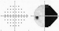
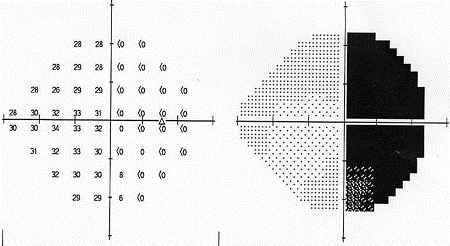
Left eye of patient with homonymous field defect
From Dr Prem S. Subramanian's personal collection; used with permission
See this image in context in the following section/s:
Evaluation of vision loss
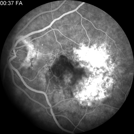
Fluorescein angiogram showing classic choroidal neovascularization with early hyperfluorescence
From Dr Prem S. Subramanian's personal collection; used with permission
See this image in context in the following section/s:
Evaluation of vision loss
Right eye of patient with homonymous field defect
From Dr Prem S. Subramanian's personal collection; used with permission
See this image in context in the following section/s:
Evaluation of vision loss
Central retinal vein occlusion: extensive retinal hemorrhages and dilated vessels
From Dr Prem S. Subramanian's personal collection; used with permission
See this image in context in the following section/s:

Evaluation of vision loss
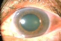
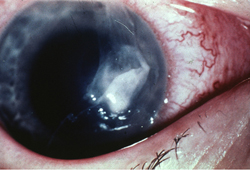
Angle-closure glaucoma: the pupil is mid-dilated, and the cornea is edematous and cloudy, as indicated by the dulled and irregular light reflex
From Dr Prem S. Subramanian's personal collection; used with permission
See this image in context in the following section/s:
Evaluation of vision loss
Corneal ulcer with epithelial defect and stromal infiltrate
From Dr Prem S. Subramanian's personal collection; used with permission
See this image in context in the following section/s:

Evaluation of vision loss
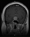
Pituitary tumor: homogenous suprasellar mass elevating and compressing optic chiasm (MRI)
From Dr Prem S. Subramanian's personal collection; used with permission
See this image in context in the following section/s:
Evaluation of vision loss
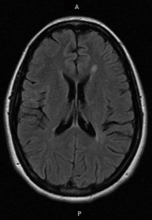
Typical white matter lesions indicative of multiple sclerosis risk (MRI)
From Dr Prem S. Subramanian's personal collection; used with permission
See this image in context in the following section/s:

Evaluation of vision loss
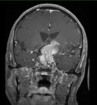
Pituitary apoplexy: large suprasellar mass with heterogeneous gadolinium enhancement (T1-weighted MRI)
From Dr Prem S. Subramanian's personal collection; used with permission
See this image in context in the following section/s:

Evaluation of vision loss
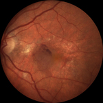
Subretinal hemorrhage and retinal elevation from subfoveal neovascularization
From Dr Prem S. Subramanian's personal collection; used with permission
See this image in context in the following section/s:

Evaluation of vision loss
Right optic nerve with disk swelling
From Dr Prem S. Subramanian's personal collection; used with permission
See this image in context in the following section/s:
Use of this content is subject to our disclaimer