Images and videos
Images


Evaluation of neck pain
Bilateral facet dislocation
With permission of the University of Colorado Department of Radiology
See this image in context in the following section/s:

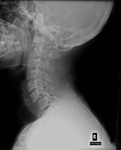
Evaluation of neck pain
Normal lateral cervical spine x-ray
With permission of the University of Colorado Department of Radiology
See this image in context in the following section/s:

Evaluation of neck pain
C3-4 diskitis with endplate destruction
With permission of the University of Colorado Department of Radiology
See this image in context in the following section/s:

Evaluation of neck pain
Zostiform vesicular eruption within the D8 dermatome on the left
BMJ Case Reports 2009; doi:10.1136/bcr.2006.114116. Copyright © 2011 by the BMJ Publishing Group Ltd
See this image in context in the following section/s:

Evaluation of neck pain
Patient with severe left torticollis (note hypertrophy of right sternocleidomastoid muscle)
From the personal collection of David B. Sommer, MD, MPH
See this image in context in the following section/s:

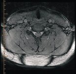
Evaluation of neck pain
MRI cervical spine. Axial three-dimensional cosmic at C5/C6 with anterior indentation of the thecal sac
BMJ Case Reports 2009; doi:10.1136/bcr.07.2008.0573. Copyright © 2011 by the BMJ Publishing Group Ltd
See this image in context in the following section/s:

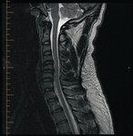
Evaluation of neck pain
MRI cervical spine. Sagittal T1, SE with a central disc at C5/C6 indenting the thecal sac
BMJ Case Reports 2009; doi:10.1136/bcr.07.2008.0573. Copyright © 2011 by the BMJ Publishing Group Ltd
See this image in context in the following section/s:
Use of this content is subject to our disclaimer