Images and videos
Images

Eosinophilic granulomatosis with polyangiitis (Churg-Strauss syndrome)
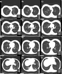
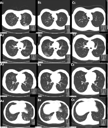
CT scan of the chest obtained (A) during symptom exacerbation, revealing pulmonary infiltrates in the right inferior lobe (A3) and the left inferior lobe (A4). Re-evaluation 15 days later (B) revealed the migratory character of the lesions, with complete disappearance of the previously described infiltrates and the presence of new areas of ground glass opacities within the right inferior lobe (B2) and the left lingular and left inferior lobe (B4). (C) Six months after treatment, all the lung lesions described had healed completely
BMJ Case Reports 2009; doi:10.1136/bcr.04.2009.1731. Copyright © 2011 by the BMJ Publishing Group Ltd
See this image in context in the following section/s:
Eosinophilic granulomatosis with polyangiitis (Churg-Strauss syndrome)
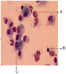
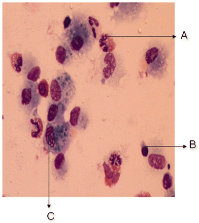
Cytological examination of the bronchoalveolar lavage fluid, revealing the presence of a vast predominance of eosinophils (A), representing 27% of the cellular elements. The other cellular elements found were macrophages (C) and lymphocytes (B)
BMJ Case Reports 2009; doi:10.1136/bcr.04.2009.1731. Copyright © 2011 by the BMJ Publishing Group Ltd
See this image in context in the following section/s:
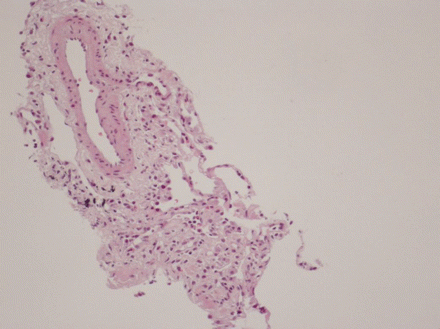
Eosinophilic granulomatosis with polyangiitis (Churg-Strauss syndrome)
Histological evaluation of the lung biopsy specimen, revealing the presence of an inflammatory infiltrate composed predominantly of eosinophils found within both the vascular lumen and the vascular wall
BMJ Case Reports 2009; doi:10.1136/bcr.04.2009.1731. Copyright © 2011 by the BMJ Publishing Group Ltd
See this image in context in the following section/s:
Use of this content is subject to our disclaimer