Images and videos
Images

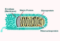


Rabies
Illustration of rabies virus in cross-section. Concentric layers: envelope membrane bilayer, M protein, and tightly coiled encased RNA
CDC
See this image in context in the following section/s:

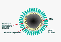
Rabies
Illustration of rabies virus in longitudinal section
CDC
See this image in context in the following section/s:
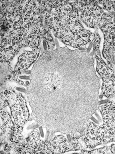
Rabies
This transmission electron micrograph reveals the presence of Lagos bat virus virions and an intracytoplasmic inclusion body in this tissue sample
CDC; Dr Fred Murphy; Sylvia Whitfield
See this image in context in the following section/s:
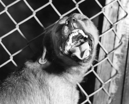
Rabies
A canine suspected of being rabid that had been exhibiting signs of restlessness and overall uncharacteristic aggressive behavior
CDC
See this image in context in the following section/s:
Use of this content is subject to our disclaimer