Large bowel obstruction should be identified as early as possible in any patient with abdominal pain and a change in bowel habit as it is a potentially life-threatening surgical emergency.[4]Association of Surgeons of Great Britain and Ireland; Royal College of Surgeons of England. Commissioning guide: emergency general surgery (acute abdominal pain). April 2014 [internet publication].
https://www.rcseng.ac.uk/library-and-publications/rcs-publications/docs/emergency-general-guide
Patients with signs of peritonitis, perforation, or with recurrent or unsuccessful nonoperative decompression should be referred immediately for surgical management.[8]Tian BWCA, Vigutto G, Tan E, et al. WSES consensus guidelines on sigmoid volvulus management. World J Emerg Surg. 2023 May 15;18(1):34.
https://wjes.biomedcentral.com/articles/10.1186/s13017-023-00502-x
http://www.ncbi.nlm.nih.gov/pubmed/37189134?tool=bestpractice.com
[13]Naveed M, Jamil LH, Fujii-Lau LL, et al. American Society for Gastrointestinal Endoscopy guideline on the role of endoscopy in the management of acute colonic pseudo-obstruction and colonic volvulus. Gastrointest Endosc. 2020 Feb;91(2):228-35.
http://www.ncbi.nlm.nih.gov/pubmed/31791596?tool=bestpractice.com
[26]Bruzzi M, Lefèvre JH, Desaint B, et al. Management of acute sigmoid volvulus: short- and long-term results. Colorectal Dis. 2015 Oct;17(10):922-8.
http://www.ncbi.nlm.nih.gov/pubmed/25808350?tool=bestpractice.com
Signs and symptoms depend on the underlying cause; therefore, a thorough physical exam is important.[3]Alavi K, Poylin V, Davids JS, et al. The American Society of Colon and Rectal Surgeons clinical practice guidelines for the management of colonic volvulus and acute colonic pseudo-obstruction. Dis Colon Rectum. 2021 Sep 1;64(9):1046-57.
https://journals.lww.com/dcrjournal/Fulltext/2021/09000/The_American_Society_of_Colon_and_Rectal_Surgeons.5.aspx
http://www.ncbi.nlm.nih.gov/pubmed/34016826?tool=bestpractice.com
The classic signs and symptoms are intermittent abdominal pain, distention, vomiting, nausea, and absolute constipation.[4]Association of Surgeons of Great Britain and Ireland; Royal College of Surgeons of England. Commissioning guide: emergency general surgery (acute abdominal pain). April 2014 [internet publication].
https://www.rcseng.ac.uk/library-and-publications/rcs-publications/docs/emergency-general-guide
Patient age and onset of symptoms (gradual versus abrupt) guides diagnosis of the underlying cause.
Initial workup involves laboratory tests and radiologic evaluation. In hemodynamically stable patients, colonic volvulus is often initially evaluated with plain abdominal radiographs.[3]Alavi K, Poylin V, Davids JS, et al. The American Society of Colon and Rectal Surgeons clinical practice guidelines for the management of colonic volvulus and acute colonic pseudo-obstruction. Dis Colon Rectum. 2021 Sep 1;64(9):1046-57.
https://journals.lww.com/dcrjournal/Fulltext/2021/09000/The_American_Society_of_Colon_and_Rectal_Surgeons.5.aspx
http://www.ncbi.nlm.nih.gov/pubmed/34016826?tool=bestpractice.com
Use abdominal plain x-ray where there is a clinical suspicion of bowel obstruction; the presence of obstruction may be confirmed by colonic dilation on plain abdominal x-ray.[5]Pisano M, Zorcolo L, Merli C, et al. 2017 WSES guidelines on colon and rectal cancer emergencies: obstruction and perforation. World J Emerg Surg. 2018 Aug 13;13:36.
https://www.ncbi.nlm.nih.gov/pmc/articles/PMC6090779
http://www.ncbi.nlm.nih.gov/pubmed/30123315?tool=bestpractice.com
Large bowel obstruction may also be diagnosed using ultrasound (performed by radiologists or as point of care), however, the diagnostic criteria are not as well defined as for small bowel obstruction.[27]Hollerweger A, Maconi G, Ripolles T, et al. Gastrointestinal ultrasound (GIUS) in intestinal emergencies - an EFSUMB position paper. Ultraschall Med. 2020 Dec;41(6):646-57.
https://www.thieme-connect.com/products/ejournals/html/10.1055/a-1147-1295
http://www.ncbi.nlm.nih.gov/pubmed/32311749?tool=bestpractice.com
[28]Li RT, Zhao Y, Zou XJ, et al. Overview of point-of-care ultrasound in diagnosing intestinal obstruction. World J Emerg Med. 2022;13(2):135-40.
https://www.ncbi.nlm.nih.gov/pmc/articles/PMC8861339
In patients where clinical assessment and plain abdominal radiographs are insufficient to confirm the diagnosis of colonic volvulus, computed tomography (CT) imaging, with or without rectal contrast, should be considered.[2]Miller AS, Boyce K, Box B, et al. The Association of Coloproctology of Great Britain and Ireland consensus guidelines in emergency colorectal surgery. Colorectal Dis. 2021 Feb;23(2):476-547.
https://onlinelibrary.wiley.com/doi/10.1111/codi.15503
http://www.ncbi.nlm.nih.gov/pubmed/33470518?tool=bestpractice.com
[3]Alavi K, Poylin V, Davids JS, et al. The American Society of Colon and Rectal Surgeons clinical practice guidelines for the management of colonic volvulus and acute colonic pseudo-obstruction. Dis Colon Rectum. 2021 Sep 1;64(9):1046-57.
https://journals.lww.com/dcrjournal/Fulltext/2021/09000/The_American_Society_of_Colon_and_Rectal_Surgeons.5.aspx
http://www.ncbi.nlm.nih.gov/pubmed/34016826?tool=bestpractice.com
[13]Naveed M, Jamil LH, Fujii-Lau LL, et al. American Society for Gastrointestinal Endoscopy guideline on the role of endoscopy in the management of acute colonic pseudo-obstruction and colonic volvulus. Gastrointest Endosc. 2020 Feb;91(2):228-35.
http://www.ncbi.nlm.nih.gov/pubmed/31791596?tool=bestpractice.com
CT scan is the best imaging technique to evaluate large bowel obstruction and perforation and may indicate the underlying cause (e.g., malignancy, colonic volvulus, stricture, diverticulitis).[3]Alavi K, Poylin V, Davids JS, et al. The American Society of Colon and Rectal Surgeons clinical practice guidelines for the management of colonic volvulus and acute colonic pseudo-obstruction. Dis Colon Rectum. 2021 Sep 1;64(9):1046-57.
https://journals.lww.com/dcrjournal/Fulltext/2021/09000/The_American_Society_of_Colon_and_Rectal_Surgeons.5.aspx
http://www.ncbi.nlm.nih.gov/pubmed/34016826?tool=bestpractice.com
[5]Pisano M, Zorcolo L, Merli C, et al. 2017 WSES guidelines on colon and rectal cancer emergencies: obstruction and perforation. World J Emerg Surg. 2018 Aug 13;13:36.
https://www.ncbi.nlm.nih.gov/pmc/articles/PMC6090779
http://www.ncbi.nlm.nih.gov/pubmed/30123315?tool=bestpractice.com
[8]Tian BWCA, Vigutto G, Tan E, et al. WSES consensus guidelines on sigmoid volvulus management. World J Emerg Surg. 2023 May 15;18(1):34.
https://wjes.biomedcentral.com/articles/10.1186/s13017-023-00502-x
http://www.ncbi.nlm.nih.gov/pubmed/37189134?tool=bestpractice.com
[29]Beattie GC, Peters RT, Guy S, et al. Computed tomography in the assessment of suspected large bowel obstruction. ANZ J Surg. 2007 Mar;77(3):160-5.
http://www.ncbi.nlm.nih.gov/pubmed/17305992?tool=bestpractice.com
Do not use a protocol that involves contrast-enhanced CT acquisition in addition to unenhanced acquisition except in certain circumstances (such as gastrointestinal hemorrhage), as unenhanced images do not add diagnostic information.[30]American College of Radiology. Ten things physicians and patients should question. Choosing Wisely, an initiative of the ABIM Foundation. 2021 [internet publication].
https://web.archive.org/web/20230330210926/https://www.choosingwisely.org/societies/american-college-of-radiology
Endoscopy and biopsy confirm a diagnosis of malignancy, although this is frequently not achievable and may be inappropriate in the emergency situation.
History
A thorough history should be noted. It may be difficult to obtain an accurate history in patients with neuropsychiatric disorders, or in patients residing in long-term care facilities who rely on others to relay key historical events.[3]Alavi K, Poylin V, Davids JS, et al. The American Society of Colon and Rectal Surgeons clinical practice guidelines for the management of colonic volvulus and acute colonic pseudo-obstruction. Dis Colon Rectum. 2021 Sep 1;64(9):1046-57.
https://journals.lww.com/dcrjournal/Fulltext/2021/09000/The_American_Society_of_Colon_and_Rectal_Surgeons.5.aspx
http://www.ncbi.nlm.nih.gov/pubmed/34016826?tool=bestpractice.com
Features common to all causes of obstruction include colicky abdominal pain and abdominal distension, altered bowel habit with either a failure to pass feces (complete obstruction) or a successful passing of some flatus or feces (partial obstruction).[5]Pisano M, Zorcolo L, Merli C, et al. 2017 WSES guidelines on colon and rectal cancer emergencies: obstruction and perforation. World J Emerg Surg. 2018 Aug 13;13:36.
https://www.ncbi.nlm.nih.gov/pmc/articles/PMC6090779
http://www.ncbi.nlm.nih.gov/pubmed/30123315?tool=bestpractice.com
Vomiting is less frequent than in small bowel obstruction.[5]Pisano M, Zorcolo L, Merli C, et al. 2017 WSES guidelines on colon and rectal cancer emergencies: obstruction and perforation. World J Emerg Surg. 2018 Aug 13;13:36.
https://www.ncbi.nlm.nih.gov/pmc/articles/PMC6090779
http://www.ncbi.nlm.nih.gov/pubmed/30123315?tool=bestpractice.com
Continuous pain may indicate bowel ischemia.[4]Association of Surgeons of Great Britain and Ireland; Royal College of Surgeons of England. Commissioning guide: emergency general surgery (acute abdominal pain). April 2014 [internet publication].
https://www.rcseng.ac.uk/library-and-publications/rcs-publications/docs/emergency-general-guide
Other more specific features due to the underlying cause are listed below.
Colorectal malignancy
Colonic obstruction occurs more commonly in older people, and is more likely to be due to an underlying colorectal malignancy in this age group.[5]Pisano M, Zorcolo L, Merli C, et al. 2017 WSES guidelines on colon and rectal cancer emergencies: obstruction and perforation. World J Emerg Surg. 2018 Aug 13;13:36.
https://www.ncbi.nlm.nih.gov/pmc/articles/PMC6090779
http://www.ncbi.nlm.nih.gov/pubmed/30123315?tool=bestpractice.com
Symptom onset may be gradual.[4]Association of Surgeons of Great Britain and Ireland; Royal College of Surgeons of England. Commissioning guide: emergency general surgery (acute abdominal pain). April 2014 [internet publication].
https://www.rcseng.ac.uk/library-and-publications/rcs-publications/docs/emergency-general-guide
Recent weight loss and rectal bleeding mixed with stools and new-onset abdominal pain suggests an underlying malignancy, especially if in conjunction with reduction change in bowel habit.[5]Pisano M, Zorcolo L, Merli C, et al. 2017 WSES guidelines on colon and rectal cancer emergencies: obstruction and perforation. World J Emerg Surg. 2018 Aug 13;13:36.
https://www.ncbi.nlm.nih.gov/pmc/articles/PMC6090779
http://www.ncbi.nlm.nih.gov/pubmed/30123315?tool=bestpractice.com
Colonic volvulus
More prevalent in institutionalized patients or patients with mental illness; patients with a prior history of colonic volvulus, megacolon, or previous abdominal surgery; patients who use laxatives chronically; and patients with diabetes.[13]Naveed M, Jamil LH, Fujii-Lau LL, et al. American Society for Gastrointestinal Endoscopy guideline on the role of endoscopy in the management of acute colonic pseudo-obstruction and colonic volvulus. Gastrointest Endosc. 2020 Feb;91(2):228-35.
http://www.ncbi.nlm.nih.gov/pubmed/31791596?tool=bestpractice.com
[21]Halabi WJ, Jafari MD, Kang CY, et al. Colonic volvulus in the United States: trends, outcomes, and predictors of mortality. Ann Surg. 2014 Feb;259(2):293-301.
http://www.ncbi.nlm.nih.gov/pubmed/23511842?tool=bestpractice.com
Although the duration of symptoms before presentation ranges from a few hours to several days, cecal volvulus tends to present more acutely, whereas sigmoid volvulus often has a more indolent presentation.[3]Alavi K, Poylin V, Davids JS, et al. The American Society of Colon and Rectal Surgeons clinical practice guidelines for the management of colonic volvulus and acute colonic pseudo-obstruction. Dis Colon Rectum. 2021 Sep 1;64(9):1046-57.
https://journals.lww.com/dcrjournal/Fulltext/2021/09000/The_American_Society_of_Colon_and_Rectal_Surgeons.5.aspx
http://www.ncbi.nlm.nih.gov/pubmed/34016826?tool=bestpractice.com
Diverticular disease
Rarely produces complete colonic obstruction.
Symptom onset is gradual.
Patient has had prior recurrent episodes of diverticulitis with abdominal pain and tenderness.
See Diverticular disease.
Benign strictures
Prior radiotherapy history for cancer in patients with radiotherapy-induced stricture. Radiotherapy usually produces effects on the sigmoid colon and dependent ileal loops. A sigmoid stricture usually occurs at the rectosigmoid and presents with abdominal pain and tenesmus due to small volumes of feces repeatedly entering the rectum.
History of aortic surgery or ischemic colitis may indicate an ischemic colonic stricture.
History of colonic resection may indicate an anastomotic stricture.
Other causes
Fever and increasing constant pain, and pain on movement, coughing, or deep breathing, may imply perforation or impending perforation. However, fever may arise from concurrent illness or be implicated in a rarer cause of obstruction such as pelvic sepsis or inflammatory bowel disease.
Physical examination
Abdominal distension in the distribution of the affected colonic segments and tympanic abdomen are common to all causes of mechanical obstruction.[3]Alavi K, Poylin V, Davids JS, et al. The American Society of Colon and Rectal Surgeons clinical practice guidelines for the management of colonic volvulus and acute colonic pseudo-obstruction. Dis Colon Rectum. 2021 Sep 1;64(9):1046-57.
https://journals.lww.com/dcrjournal/Fulltext/2021/09000/The_American_Society_of_Colon_and_Rectal_Surgeons.5.aspx
http://www.ncbi.nlm.nih.gov/pubmed/34016826?tool=bestpractice.com
[5]Pisano M, Zorcolo L, Merli C, et al. 2017 WSES guidelines on colon and rectal cancer emergencies: obstruction and perforation. World J Emerg Surg. 2018 Aug 13;13:36.
https://www.ncbi.nlm.nih.gov/pmc/articles/PMC6090779
http://www.ncbi.nlm.nih.gov/pubmed/30123315?tool=bestpractice.com
Mild tenderness may be present in early obstruction from any cause. Fever may indicate an urgent complication of bowel obstruction, such as septic shock or impending perforation.[5]Pisano M, Zorcolo L, Merli C, et al. 2017 WSES guidelines on colon and rectal cancer emergencies: obstruction and perforation. World J Emerg Surg. 2018 Aug 13;13:36.
https://www.ncbi.nlm.nih.gov/pmc/articles/PMC6090779
http://www.ncbi.nlm.nih.gov/pubmed/30123315?tool=bestpractice.com
Severe tenderness and abdominal rigidity imply peritonitis secondary to perforation. Tenderness, particularly over the cecum, could indicate impending perforation.[4]Association of Surgeons of Great Britain and Ireland; Royal College of Surgeons of England. Commissioning guide: emergency general surgery (acute abdominal pain). April 2014 [internet publication].
https://www.rcseng.ac.uk/library-and-publications/rcs-publications/docs/emergency-general-guide
Bowel sounds may be normal initially, but become quiet as obstruction progresses. Digital rectal exam can be used to diagnose low rectal cancer and assess rectal contents.[5]Pisano M, Zorcolo L, Merli C, et al. 2017 WSES guidelines on colon and rectal cancer emergencies: obstruction and perforation. World J Emerg Surg. 2018 Aug 13;13:36.
https://www.ncbi.nlm.nih.gov/pmc/articles/PMC6090779
http://www.ncbi.nlm.nih.gov/pubmed/30123315?tool=bestpractice.com
An empty vault suggests complete obstruction above the reach of the examining finger. This finding is usually seen in sigmoid and cecal volvulus.[3]Alavi K, Poylin V, Davids JS, et al. The American Society of Colon and Rectal Surgeons clinical practice guidelines for the management of colonic volvulus and acute colonic pseudo-obstruction. Dis Colon Rectum. 2021 Sep 1;64(9):1046-57.
https://journals.lww.com/dcrjournal/Fulltext/2021/09000/The_American_Society_of_Colon_and_Rectal_Surgeons.5.aspx
http://www.ncbi.nlm.nih.gov/pubmed/34016826?tool=bestpractice.com
Hard stools imply fecal impaction. Low anastomosis or rectal masses can be palpated during digital rectal exam. Digital rectal exam may also identify a pelvic mass suggestive of a gynecologic malignancy or abscess and may identify a foreign body. In women, pelvic exam may detect a mass suggestive of a gynecologic malignancy or abscess. Irreducible hernias should be excluded, and previous incisions and the groin area should be carefully inspected.
A positive fecal occult blood test result mandates exclusion of a more proximal large bowel malignancy.
Laboratory investigations
Serum amylase/lipase, complete blood count, serum electrolytes, renal function, and coagulation studies should form part of the initial assessment.[4]Association of Surgeons of Great Britain and Ireland; Royal College of Surgeons of England. Commissioning guide: emergency general surgery (acute abdominal pain). April 2014 [internet publication].
https://www.rcseng.ac.uk/library-and-publications/rcs-publications/docs/emergency-general-guide
It is important to check blood glucose levels because diabetic ketoacidosis (DKA) can present with abdominal pain.[31]Umpierrez G, Freire AX. Abdominal pain in patients with hyperglycemic crises. J Crit Care. 2002 Mar;17(1):63-7.
http://www.ncbi.nlm.nih.gov/pubmed/12040551?tool=bestpractice.com
An arterial blood gas should be requested in all critically sick patients to obtain a lactate reading, which if elevated may indicate perforation or necrosis.[5]Pisano M, Zorcolo L, Merli C, et al. 2017 WSES guidelines on colon and rectal cancer emergencies: obstruction and perforation. World J Emerg Surg. 2018 Aug 13;13:36.
https://www.ncbi.nlm.nih.gov/pmc/articles/PMC6090779
http://www.ncbi.nlm.nih.gov/pubmed/30123315?tool=bestpractice.com
Urine or serum beta-human chorionic gonadotrophin (hCG) should be performed in in women of childbearing age.[32]Royal College of Surgeons of England. Emergency general surgery-commissioning guide. 2014 [internet publication].
https://www.rcseng.ac.uk/library-and-publications/rcs-publications/docs/emergency-general-guide
Tumor markers may be useful for identifying an ovarian or colonic malignancy, but results may not be available as quickly as other routine tests and are unlikely to influence initial management.
Urinalysis should be performed in anyone with urinary symptoms, and to check for urinary ketones if DKA is suspected.[31]Umpierrez G, Freire AX. Abdominal pain in patients with hyperglycemic crises. J Crit Care. 2002 Mar;17(1):63-7.
http://www.ncbi.nlm.nih.gov/pubmed/12040551?tool=bestpractice.com
[32]Royal College of Surgeons of England. Emergency general surgery-commissioning guide. 2014 [internet publication].
https://www.rcseng.ac.uk/library-and-publications/rcs-publications/docs/emergency-general-guide
X-rays
Abdominal plain x-ray is a screening test in bowel obstruction and is often used initially for diagnosis.[3]Alavi K, Poylin V, Davids JS, et al. The American Society of Colon and Rectal Surgeons clinical practice guidelines for the management of colonic volvulus and acute colonic pseudo-obstruction. Dis Colon Rectum. 2021 Sep 1;64(9):1046-57.
https://journals.lww.com/dcrjournal/Fulltext/2021/09000/The_American_Society_of_Colon_and_Rectal_Surgeons.5.aspx
http://www.ncbi.nlm.nih.gov/pubmed/34016826?tool=bestpractice.com
[5]Pisano M, Zorcolo L, Merli C, et al. 2017 WSES guidelines on colon and rectal cancer emergencies: obstruction and perforation. World J Emerg Surg. 2018 Aug 13;13:36.
https://www.ncbi.nlm.nih.gov/pmc/articles/PMC6090779
http://www.ncbi.nlm.nih.gov/pubmed/30123315?tool=bestpractice.com
An x-ray may show the colon distended to the point of the obstruction with a paucity of distal gas or signs more classically associated with colonic volvulus. Approximately 75% of colonic volvulus can be diagnosed on x-ray.[33]Mutch MG, Birnbaum EH, Menias CO. Diagnostic evaluations-radiology, nuclear scans, PET, CT colography. In: Wolff BG, Fleshman JW, Beck DE, et al, eds. The ASCRS textbook of colon and rectal surgery, 1st ed. New York, NY: Springer; 2007:57-100.[34]Jaffe T, Thompson WM. Large-bowel obstruction in the adult: classic radiographic and CT findings, etiology, and mimics. Radiology. 2015 Jun;275(3):651-63.
https://www.doi.org/10.1148/radiol.2015140916
http://www.ncbi.nlm.nih.gov/pubmed/25997131?tool=bestpractice.com
Sigmoid volvulus shows a dilated inverted U-shaped loop of colon (resembling a coffee bean or bent inner tube) projecting toward the right side of the abdomen.[8]Tian BWCA, Vigutto G, Tan E, et al. WSES consensus guidelines on sigmoid volvulus management. World J Emerg Surg. 2023 May 15;18(1):34.
https://wjes.biomedcentral.com/articles/10.1186/s13017-023-00502-x
http://www.ncbi.nlm.nih.gov/pubmed/37189134?tool=bestpractice.com
Opposing colonic walls produce a radio-opaque line. Proximal large and small bowel dilation may also be evident. In cecal volvulus, dilated right colon rotates to the left side and dilated small bowel may also be present.
[Figure caption and citation for the preceding image starts]: Large bowel obstruction. Plain radiograph showing distended large bowel loops. Note grossly dilated caecum (white arrow)Musson RE, Bickle I, Vijay RKP, et al. Gas patterns on plain abdominal radiographs: a pictorial review. Postgrad Med J. 2011 Apr;87(1026):274-87; used with permission [Citation ends]. [Figure caption and citation for the preceding image starts]: Abdominal radiograph showing dilated large bowel loops. Typical "coffee bean’"sign seen for sigmoid volvulusRoy SP, Tay YK, Kozman D. Very rare case of synchronous volvulus of the sigmoid colon and caecum causing large-bowel obstruction. BMJ Case Rep. 2019 Jan 28;12(1):bcr-2018-227375; used with permission [Citation ends].
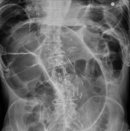
Plain abdominal x-ray is not an accurate method for confirming a malignant etiology for the obstruction. In patients with an incompetent ileocecal valve, an x-ray may also show a distended small bowel with air-fluid levels suggestive of small bowel obstruction.[3]Alavi K, Poylin V, Davids JS, et al. The American Society of Colon and Rectal Surgeons clinical practice guidelines for the management of colonic volvulus and acute colonic pseudo-obstruction. Dis Colon Rectum. 2021 Sep 1;64(9):1046-57.
https://journals.lww.com/dcrjournal/Fulltext/2021/09000/The_American_Society_of_Colon_and_Rectal_Surgeons.5.aspx
http://www.ncbi.nlm.nih.gov/pubmed/34016826?tool=bestpractice.com
Foreign body ingestion may also be identified on plain abdominal x-ray. One of the limitations of abdominal plain x-ray is the risk of false negatives of pneumoperitoneum when a small amount of intraperitoneal free air is present (e.g., in the case of early perforation at the tumor site).[5]Pisano M, Zorcolo L, Merli C, et al. 2017 WSES guidelines on colon and rectal cancer emergencies: obstruction and perforation. World J Emerg Surg. 2018 Aug 13;13:36.
https://www.ncbi.nlm.nih.gov/pmc/articles/PMC6090779
http://www.ncbi.nlm.nih.gov/pubmed/30123315?tool=bestpractice.com
Ultrasound
Ultrasound has been shown to have a comparable accuracy to CT scans in the detection of small bowel obstruction but diagnostic criteria and accuracy for large bowel obstruction are less well defined.[27]Hollerweger A, Maconi G, Ripolles T, et al. Gastrointestinal ultrasound (GIUS) in intestinal emergencies - an EFSUMB position paper. Ultraschall Med. 2020 Dec;41(6):646-57.
https://www.thieme-connect.com/products/ejournals/html/10.1055/a-1147-1295
http://www.ncbi.nlm.nih.gov/pubmed/32311749?tool=bestpractice.com
One proposed criteria include dilated large intestine (>4.5 cm) and presence of abdominal A-lines (gastrointestinal intraluminal air).[28]Li RT, Zhao Y, Zou XJ, et al. Overview of point-of-care ultrasound in diagnosing intestinal obstruction. World J Emerg Med. 2022;13(2):135-40.
https://www.ncbi.nlm.nih.gov/pmc/articles/PMC8861339
Point-of-care ultrasound (POCUS) also has the advantage that it can be performed by nonradiologists at the bedside as a noninvasive diagnostic tool without radiation but does require specialized training.
CT abdomen and pelvis
CT can confirm large bowel obstruction in >90% of cases and may reveal the underlying cause.[29]Beattie GC, Peters RT, Guy S, et al. Computed tomography in the assessment of suspected large bowel obstruction. ANZ J Surg. 2007 Mar;77(3):160-5.
http://www.ncbi.nlm.nih.gov/pubmed/17305992?tool=bestpractice.com
[35]Frager D, Rovno HD, Baer JW, et al. Prospective evaluation of colonic obstruction with computed tomography. Abdom Imaging. 1998 Mar-Apr;23(2):141-6.
http://www.ncbi.nlm.nih.gov/pubmed/9516501?tool=bestpractice.com
[36]Verheyden C, Orliac C, Millet I, et al. Large-bowel obstruction: CT findings, pitfalls, tips and tricks. Eur J Radiol. 2020 Sep;130:109155.
http://www.ncbi.nlm.nih.gov/pubmed/32711335?tool=bestpractice.com
It is noninvasive, easily obtainable, accurate, and can identify coincidental pathology that may otherwise be missed with plain radiographs or fluoroscopic contrast studies.[3]Alavi K, Poylin V, Davids JS, et al. The American Society of Colon and Rectal Surgeons clinical practice guidelines for the management of colonic volvulus and acute colonic pseudo-obstruction. Dis Colon Rectum. 2021 Sep 1;64(9):1046-57.
https://journals.lww.com/dcrjournal/Fulltext/2021/09000/The_American_Society_of_Colon_and_Rectal_Surgeons.5.aspx
http://www.ncbi.nlm.nih.gov/pubmed/34016826?tool=bestpractice.com
CT is the preferred confirmatory diagnostic study for both cecal and sigmoid volvulus.[3]Alavi K, Poylin V, Davids JS, et al. The American Society of Colon and Rectal Surgeons clinical practice guidelines for the management of colonic volvulus and acute colonic pseudo-obstruction. Dis Colon Rectum. 2021 Sep 1;64(9):1046-57.
https://journals.lww.com/dcrjournal/Fulltext/2021/09000/The_American_Society_of_Colon_and_Rectal_Surgeons.5.aspx
http://www.ncbi.nlm.nih.gov/pubmed/34016826?tool=bestpractice.com
[8]Tian BWCA, Vigutto G, Tan E, et al. WSES consensus guidelines on sigmoid volvulus management. World J Emerg Surg. 2023 May 15;18(1):34.
https://wjes.biomedcentral.com/articles/10.1186/s13017-023-00502-x
http://www.ncbi.nlm.nih.gov/pubmed/37189134?tool=bestpractice.com
[13]Naveed M, Jamil LH, Fujii-Lau LL, et al. American Society for Gastrointestinal Endoscopy guideline on the role of endoscopy in the management of acute colonic pseudo-obstruction and colonic volvulus. Gastrointest Endosc. 2020 Feb;91(2):228-35.
http://www.ncbi.nlm.nih.gov/pubmed/31791596?tool=bestpractice.com
CT with multiplanar reconstruction can diagnose volvulus with near 100% sensitivity and 90% specificity.[13]Naveed M, Jamil LH, Fujii-Lau LL, et al. American Society for Gastrointestinal Endoscopy guideline on the role of endoscopy in the management of acute colonic pseudo-obstruction and colonic volvulus. Gastrointest Endosc. 2020 Feb;91(2):228-35.
http://www.ncbi.nlm.nih.gov/pubmed/31791596?tool=bestpractice.com
Additionally, CT can detect local and regional metastases in patients with large bowel obstruction secondary to malignancy, and may obviate the requirement for contrast enema where colorectal or pelvic/gynecologic pathology is clearly identifiable.[3]Alavi K, Poylin V, Davids JS, et al. The American Society of Colon and Rectal Surgeons clinical practice guidelines for the management of colonic volvulus and acute colonic pseudo-obstruction. Dis Colon Rectum. 2021 Sep 1;64(9):1046-57.
https://journals.lww.com/dcrjournal/Fulltext/2021/09000/The_American_Society_of_Colon_and_Rectal_Surgeons.5.aspx
http://www.ncbi.nlm.nih.gov/pubmed/34016826?tool=bestpractice.com
If colorectal pathology is apparent on CT imaging, close liaison with a radiologist is required to determine whether a contrast enema is needed, for example, to clarify whether a stricture is benign or malignant.
CT with intravenous contrast, with or without contrast enema, may be appropriate if there is uncertainty about the site of the obstruction.[5]Pisano M, Zorcolo L, Merli C, et al. 2017 WSES guidelines on colon and rectal cancer emergencies: obstruction and perforation. World J Emerg Surg. 2018 Aug 13;13:36.
https://www.ncbi.nlm.nih.gov/pmc/articles/PMC6090779
http://www.ncbi.nlm.nih.gov/pubmed/30123315?tool=bestpractice.com
Do not use a protocol that involves contrast-enhanced CT acquisition in addition to unenhanced acquisition except in certain circumstances (such as gastrointestinal hemorrhage), as unenhanced images do not add diagnostic information.[30]American College of Radiology. Ten things physicians and patients should question. Choosing Wisely, an initiative of the ABIM Foundation. 2021 [internet publication].
https://web.archive.org/web/20230330210926/https://www.choosingwisely.org/societies/american-college-of-radiology
In case of a perforation diagnosis on abdominal x-ray in a stable patient, an abdominal CT scan could be considered in order to define the cause and site of perforation. A CT scan, however, should not delay appropriate treatment. Early involvement of the surgeon is required to help make the decision to proceed with CT.[5]Pisano M, Zorcolo L, Merli C, et al. 2017 WSES guidelines on colon and rectal cancer emergencies: obstruction and perforation. World J Emerg Surg. 2018 Aug 13;13:36.
https://www.ncbi.nlm.nih.gov/pmc/articles/PMC6090779
http://www.ncbi.nlm.nih.gov/pubmed/30123315?tool=bestpractice.com
Contrast enema
May be performed after the initial assessment to confirm diagnosis, although a CT scan is typically the study of choice and is more readily available. Contrast enema provides information on the level, degree, and type of obstruction.[5]Pisano M, Zorcolo L, Merli C, et al. 2017 WSES guidelines on colon and rectal cancer emergencies: obstruction and perforation. World J Emerg Surg. 2018 Aug 13;13:36.
https://www.ncbi.nlm.nih.gov/pmc/articles/PMC6090779
http://www.ncbi.nlm.nih.gov/pubmed/30123315?tool=bestpractice.com
Water-soluble contrast flows freely to the obstruction, where there is a characteristic cut-off point depending on the nature of the obstruction. In the setting of sigmoid colonic volvulus, a smooth, tapered "bird's beak" is found at the rectosigmoid junction.[3]Alavi K, Poylin V, Davids JS, et al. The American Society of Colon and Rectal Surgeons clinical practice guidelines for the management of colonic volvulus and acute colonic pseudo-obstruction. Dis Colon Rectum. 2021 Sep 1;64(9):1046-57.
https://journals.lww.com/dcrjournal/Fulltext/2021/09000/The_American_Society_of_Colon_and_Rectal_Surgeons.5.aspx
http://www.ncbi.nlm.nih.gov/pubmed/34016826?tool=bestpractice.com
Water-soluble medium is preferred over barium, as it does not produce chemical peritonitis if there is a colonic perforation. By avoiding barium, subsequent radiologic studies are also not compromised. Also, if fecal impaction is identified, water-soluble contrast can be both diagnostic and therapeutic.
Endoscopy
Endoscopy can be both diagnostic and therapeutic.[13]Naveed M, Jamil LH, Fujii-Lau LL, et al. American Society for Gastrointestinal Endoscopy guideline on the role of endoscopy in the management of acute colonic pseudo-obstruction and colonic volvulus. Gastrointest Endosc. 2020 Feb;91(2):228-35.
http://www.ncbi.nlm.nih.gov/pubmed/31791596?tool=bestpractice.com
For patients with uncomplicated sigmoid volvulus, the American Society for Gastrointestinal Endoscopy and World Society or Emergency Surgery suggests endoscopy as the initial treatment modality.[8]Tian BWCA, Vigutto G, Tan E, et al. WSES consensus guidelines on sigmoid volvulus management. World J Emerg Surg. 2023 May 15;18(1):34.
https://wjes.biomedcentral.com/articles/10.1186/s13017-023-00502-x
http://www.ncbi.nlm.nih.gov/pubmed/37189134?tool=bestpractice.com
[13]Naveed M, Jamil LH, Fujii-Lau LL, et al. American Society for Gastrointestinal Endoscopy guideline on the role of endoscopy in the management of acute colonic pseudo-obstruction and colonic volvulus. Gastrointest Endosc. 2020 Feb;91(2):228-35.
http://www.ncbi.nlm.nih.gov/pubmed/31791596?tool=bestpractice.com
For obstructing malignant lesions in the left and rectosigmoid colon, flexible endoscopy can confirm the diagnosis based on endoscopic features and with biopsy, and may also have a therapeutic role if the lesion is amenable to stenting or dilation.[37]ASGE Standards of Practice Committee, Fisher DA, Shergill AK, et al. Role of endoscopy in the staging and management of colorectal cancer. Gastrointest Endosc. 2013 Jul;78(1):8-12.
https://www.giejournal.org/article/S0016-5107(13)01779-3/fulltext
[38]ASGE Standards of Practice Committee, Harrison ME, Anderson MA, et al. The role of endoscopy in the management of patients with known and suspected colonic obstruction and pseudo-obstruction. Gastrointest Endosc. 2010 Apr;71(4):669-79.
https://www.giejournal.org/article/S0016-5107(13)01779-3/fulltext
[39]Beck DE. Endoscopic management of bowel obstruction. Clin Colon Rectal Surg. 2021 Jul;34(4):262-8.
https://www.ncbi.nlm.nih.gov/pmc/articles/PMC8292002
http://www.ncbi.nlm.nih.gov/pubmed/34305475?tool=bestpractice.com
Endoscopy, however is associated with adverse events including perforation. The pooled risk of perforation during colonoscopy is 5.8 per 10,000 colonoscopies and is likely higher if there is a therapeutic intervention.[40]Kothari ST, Huang RJ, Shaukat A, et al. ASGE review of adverse events in colonoscopy. Gastrointest Endosc. 2019 Dec;90(6):863-76.e33.
https://www.giejournal.org/article/S0016-5107(19)32115-7/fulltext
http://www.ncbi.nlm.nih.gov/pubmed/31563271?tool=bestpractice.com
For patients with uncomplicated sigmoid volvulus, the American Society for Gastrointestinal Endoscopy and World Society of Emergency Surgery suggest endoscopy as the initial treatment modality.[8]Tian BWCA, Vigutto G, Tan E, et al. WSES consensus guidelines on sigmoid volvulus management. World J Emerg Surg. 2023 May 15;18(1):34.
https://wjes.biomedcentral.com/articles/10.1186/s13017-023-00502-x
http://www.ncbi.nlm.nih.gov/pubmed/37189134?tool=bestpractice.com
[13]Naveed M, Jamil LH, Fujii-Lau LL, et al. American Society for Gastrointestinal Endoscopy guideline on the role of endoscopy in the management of acute colonic pseudo-obstruction and colonic volvulus. Gastrointest Endosc. 2020 Feb;91(2):228-35.
http://www.ncbi.nlm.nih.gov/pubmed/31791596?tool=bestpractice.com
In sigmoid volvulus, endoscopy may visualize the classic mucosal pinwheel at the point of obstruction before detorsion.[3]Alavi K, Poylin V, Davids JS, et al. The American Society of Colon and Rectal Surgeons clinical practice guidelines for the management of colonic volvulus and acute colonic pseudo-obstruction. Dis Colon Rectum. 2021 Sep 1;64(9):1046-57.
https://journals.lww.com/dcrjournal/Fulltext/2021/09000/The_American_Society_of_Colon_and_Rectal_Surgeons.5.aspx
http://www.ncbi.nlm.nih.gov/pubmed/34016826?tool=bestpractice.com
[8]Tian BWCA, Vigutto G, Tan E, et al. WSES consensus guidelines on sigmoid volvulus management. World J Emerg Surg. 2023 May 15;18(1):34.
https://wjes.biomedcentral.com/articles/10.1186/s13017-023-00502-x
http://www.ncbi.nlm.nih.gov/pubmed/37189134?tool=bestpractice.com
Safe reduction of a sigmoid volvulus by flexible endoscopy (flexible sigmoidoscopy/colonoscopy) allows concurrent assessment of the colonic mucosa to ensure viability.[3]Alavi K, Poylin V, Davids JS, et al. The American Society of Colon and Rectal Surgeons clinical practice guidelines for the management of colonic volvulus and acute colonic pseudo-obstruction. Dis Colon Rectum. 2021 Sep 1;64(9):1046-57.
https://journals.lww.com/dcrjournal/Fulltext/2021/09000/The_American_Society_of_Colon_and_Rectal_Surgeons.5.aspx
http://www.ncbi.nlm.nih.gov/pubmed/34016826?tool=bestpractice.com
Rigid sigmoidoscopy produces the same effect but inspection of the colonic mucosa is difficult given the dramatic expulsion of gas and liquid feces. Blind passage of a rectal tube can be successful, but does not assess the mucosa. The colonic mucosa should be visualized before attempting detorsion.
Higher strictures not found on digital rectal exam can be identified by endoscopy.
