Images and videos
Images


Evaluation of proteinuria
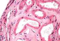
Acute tubular necrosis: biopsy showing focal areas of proximal tubule vacuolization and flattening; tubular dilation; brush border debris present in some tubular lumen
Courtesy of Puigvert Foundation, Barcelona, Spain
See this image in context in the following section/s:
Evaluation of proteinuria
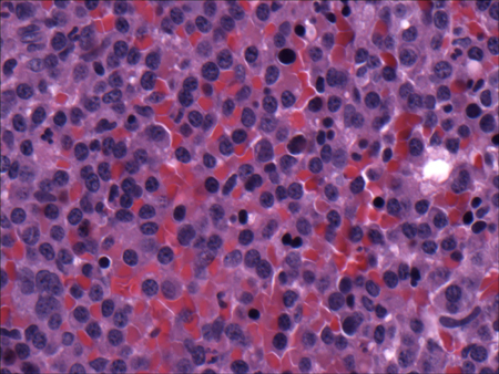
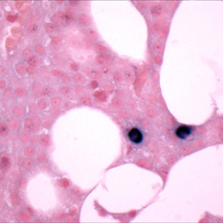
Multiple myeloma: bone marrow biopsy
Courtesy of Dr Robert Hasserjian, Hematopathology, MGH
See this image in context in the following section/s:
Evaluation of proteinuria
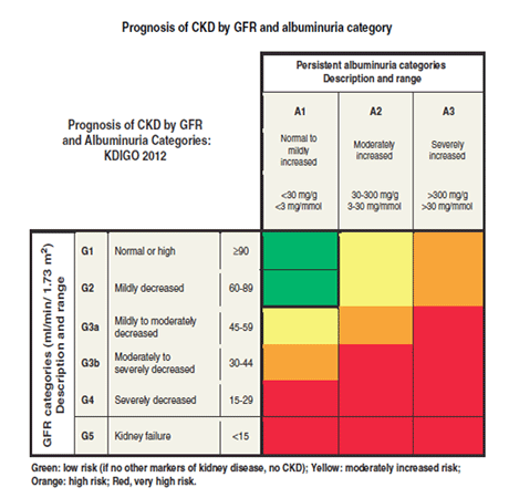
Prognosis of CKD by GFR and albuminuria category: CKD, chronic kidney disease; GFR, glomerular filtration rate; KDIGO, Kidney Disease Improving Global Outcomes
Reprinted by permission from Macmillan Publishers Ltd: Kidney International Supplements (vol 3, issue 1, January 2013), copyright 2013
See this image in context in the following section/s:
Evaluation of proteinuria
Multiple myeloma: bone marrow biopsy after histochemical analysis for lambda light chain
Courtesy of Dr Robert Hasserjian, Hematopathology, MGH
See this image in context in the following section/s:
Evaluation of proteinuria
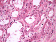
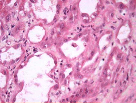
Acute tubular necrosis: biopsy showing denuded basement membranes and presence of cells in the tubule lumen
Courtesy of Puigvert Foundation, Barcelona, Spain
See this image in context in the following section/s:
Evaluation of proteinuria
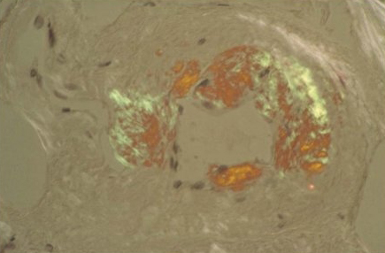
Amyloidosis: Congo red stained blood vessel in a bone marrow biopsy demonstrating pathognomonic green birefringence
Courtesy of Dr Morie A. Gertz, Hematology, Mayo Clinic, Rochester, MN
See this image in context in the following section/s:
Use of this content is subject to our disclaimer