Tests
1st tests to order
dual-energy x-ray absorptiometry (DXA)-bone mineral density (BMD)
Test
DXA is considered the gold standard for measurement of bone density; the World Health Organization (WHO) considers BMD measurements diagnostic for osteoporosis.[3][31][69][77]
It is the preferred procedure for diagnosis in many situations, although it may not be required in all circumstances. For instance, in the context of assessment for prevention of osteoporotic fractures, guidance from the UK-based National Institute for Health and Care Excellence states that for women ages 75 years or older who have one or more independent clinical risk factors for fracture or indicators of low BMD, a DXA scan may not be required if the responsible clinician considers it to be clinically inappropriate or unfeasible.[71]
T-scores are determined by measuring bone density at the hip, so are best for predicting hip fracture. Accuracy of DXA at the hip is >90%. Extrapolation of T-scores to other sites may not be as predictive of fracture.[3]
Risk of hip fracture is similar for men and women with similar BMD measurements, and it is acceptable to use the same categories of T-scores for diagnosing osteoporosis in men and women.[82] Relative risk of hip fracture is 2.6 (95% CI 2.0 to 3.5) for each standard deviation below the mean BMD for young, healthy white women.[3]
Spine DXA in older men and women may be misleading due to artifacts (e.g., aortic calcification, osteophytes).
Some studies have shown sex differences in the relationship between BMD and fracture risk. This relationship is more distinctive in younger than in older men.[85]
Result
T-score ≤-2.5 indicates osteoporosis; T-score ≤-2.5 with fragility fracture(s) indicates severe (or established) osteoporosis
Tests to consider
Fracture Risk Assessment Tool (FRAX)
Test
FRAX was developed by the WHO to assess fracture risk. FRAX integrates clinical risk factors for fracture and bone mineral density scores at the femoral neck to calculate a 10-year fracture probability for men and women.
FRAX can be used to help assess the need for BMD testing or pharmacologic treatment.[24][31][81] However, the American College of Obstetric and Gynecology highlights that the FRAX tool may be limited due to the inability to input specific amounts, dosage, or duration for alcohol intake, corticosteroid use, smoking, or the number of prior fractures. History of recent falls and spine BMD are not incorporated into the model, both of which increase the risk of osteoporotic fracture, which may lead to an underestimation of risk for these patients.[31] The FRAXplus calculator addresses some of these limitations with the introduction of additional variables (e.g., recency of fragility fracture, duration of type 2 diabetes mellitus, number of falls in the past year) to provide a more accurate estimate of fracture risk.[82]
The American College of Physicians (ACP) notes that many risk assessment tools are available, but suggests an individualized assessment of baseline fracture risk based on bone density, history of fractures, response to prior osteoporosis treatment, and other risk factors.[67]
Result
interpretation of FRAX scores varies depending on location; check local guidelines for the 10-year fracture probability percentage that triggers treatment
vertebral fracture assessment (DXA-VFA)
Test
Vertebral fracture assessment (DXA-VFA) can be performed during the same session as DXA-BMD analysis to provide images of the lumbar spine. DXA-VFA has moderate sensitivity and high specificity for detecting vertebral fractures compared with spinal radiography, with the advantages of reduced radiation exposure, lower cost, and greater convenience.[62][73][74]
In general, vertebral imaging (with either DXA-VFA or x-ray) should be considered for postmenopausal women and older men with low BMD, height loss, kyphosis, recent or ongoing long-term glucocorticoid treatment, or patients with acute onset back pain and risk factors for osteoporosis.[24][62] However, recommended indications for vertebral imaging vary; consult local guidance.
Result
may detect vertebral fracture
trabecular bone score
Test
Trabecular bone score (TBS) is a measure of bone microarchitecture derived from lumbar spine DXA images. TBS is an independent predictor of incident fracture and can be used in conjunction with BMD, clinical risk factors, and/or the FRAX to enhance the accuracy of fracture risk predictions.[69][75] It may be most useful for patients whose BMD T-score for FRAX score is close to an intervention threshold.[69][75]
Result
low score indicates osteoporotic bone at increased risk for fracture
quantitative ultrasound (QUS) of the heel
Test
QUS may be as predictive of hip fracture as dual-energy x-ray absorptiometry (DXA). However, most therapeutic studies were based on DXA.[2]
QUS may be most useful in regions without access to DXA, but the same diagnostic criteria defined for DXA cannot be applied to this technique.[3][76][77] QUS is, rather than a method for assessing bone mineral density, a method to measure unknown elements of bone strength. QUS may be a good predictor of population-based fracture, but not individual fractures. Additionally, T-score has been tested only for DXA and cannot be applied to QUS.
Result
standard parameters for diagnosing osteoporosis by QUS have not yet been determined
x-ray (wrist, heel, spine, and hip)
Test
X-ray may reveal osteopenia and/or fractures (e.g., vertebral fractures), but does not diagnose osteoporosis.[78] It is used to drive the need for dual-energy x-ray absorptiometry assessment when osteopenia is detected coincidentally. In general, vertebral imaging (with either DXA-VFA or x-ray) should be considered for postmenopausal women and older men with low BMD, height loss, kyphosis, recent or ongoing long-term glucocorticoid treatment, or patients with acute onset back pain and risk factors for osteoporosis.[24][62] However, recommended indications for vertebral imaging vary; consult local guidance.
X-ray is also used in therapeutic trials to assess fracture incidence.[78][Figure caption and citation for the preceding image starts]: Chest x-ray showing marked deformity and volume loss of bony thorax in a patient with osteoporosisBMJ Case Reports 2009; doi:10.1136/bcr.07.2008.0359. Copyright ©BMJ publishing group 2010 [Citation ends].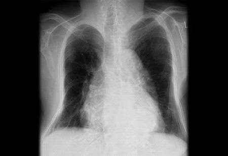
Result
may reveal osteopenia and/or fractures (e.g., vertebral fractures)
quantitative CT
Test
Alternative modality if dual-energy x-ray absorptiometry not available.[69][79][80] Quantitative CT may also be useful for assessing patients with severe degenerative spine disease, obesity, or very tall or short patients.[69]
Projectional quantitative CT can provide hip BMD measurements equivalent to DXA that may be interpreted using WHO T-score criteria, but WHO T-score criteria cannot be applied to spine quantitative CT measurements.[69][81]
Result
reduced trabecular bone density
biochemical markers of bone resorption and bone formation
Test
There is no reference range for urinary deoxypyridinoline and N-telopeptides that is diagnostic of osteoporosis.
Can be used to monitor improvement. However, a repeat dual-energy x-ray absorptiometry would be the preferred test.
Not accurate for diagnosis of osteoporosis, but, in conjunction with bone mineral density, can be useful predictors of fracture risk.[3] Bone turnover markers can be used specifically in monitoring response to treatment rather than for the approach to treatment.[83][84] It has been recommended that serum procollagen type I N propeptide (PINP) be used as a marker of bone formation, and serum C-terminal telopeptide of type I collagen (CTX) as a reference of bone turnover markers.[84] However, there are constraints with their use in the US due to problems with reimbursement by insurance companies.
Result
elevated urinary deoxypyridinoline and N-telopeptides; low serum procollagen type I N propeptide and elevated serum C-terminal telopeptide of type I collagen
serum alkaline phosphatase
Test
If elevated could indicate osteomalacia.[3]
Result
normal
serum calcium
Test
Hypocalcemia could indicate osteomalacia, hypercalcemia could indicate hyperparathyroidism.[3]
Result
normal
serum albumin
Test
A marker of nutritional status that is necessary for calculating a corrected serum calcium concentration.
Result
possibly decreased
serum creatinine
Test
Necessary for estimating glomerular filtration rate and subsequent renal function with concern for chronic kidney disease (CKD)-bone and mineral disorder. In general, bone disease in CKD patients (stages 1-3) with serum PTH concentrations between 65 and 300 picograms/mL is likely osteoporosis.[86]
Result
normal
serum phosphate
Test
Low serum phosphate levels could indicate osteomalacia.[3]
Result
normal
serum 25-hydroxy vitamin D
Test
To rule out vitamin deficiency (defined as 25-hydroxyvitamin D levels of <20 nanograms/mL).[3]
Result
normal
serum parathyroid hormone
Test
Screening for hyperparathyroidism.[3]
Result
may be normal or elevated
thyroid function tests
Test
Should be performed in patients with symptomatic hyperthyroidism. Patients on a high suppressive dose with thyroid supplementation should also be tested.[3]
Decreased thyroid-stimulating hormone and elevated levels of T4 and/or T3 could indicate hyperthyroidism.
Result
normal
urinary free cortisol
Test
Subtle Cushing syndrome may occur rarely. Urinary free cortisol must be considered in those with typical phenotypic features of Cushing syndrome.
If elevated could indicate Cushing syndrome.[3]
Result
normal
serum testosterone (men)
Test
Should be considered in all young males with osteoporosis.
Decreased in cases of hypogonadism.[3]
Result
normal
urine protein electrophoresis
Test
Should be performed in older patients with multiple spontaneous vertebral fractures associated with bone pain and anemia to rule out myeloma.[3]
Result
normal
serum protein electrophoresis
Test
Should be performed in older patients with multiple spontaneous vertebral fractures associated with bone pain and anemia to rule out myeloma.[3]
Result
normal
Use of this content is subject to our disclaimer