Diagnosis is clinical, supported by testing when required. Patients should be queried about the frequency, severity, timing (day time or night time), and duration of symptoms and the presence of any specific triggers (dietary or nondietary).[5]Fass R. Gastroesophageal Reflux Disease. N Engl J Med. 2022 Sep 29;387(13):1207-16.
http://www.ncbi.nlm.nih.gov/pubmed/36170502?tool=bestpractice.com
Heartburn and regurgitation are the most reliable symptoms.[1]Katz PO, Dunbar KB, Schnoll-Sussman FH, et al. ACG clinical guideline for the diagnosis and management of gastroesophageal reflux disease. Am J Gastroenterol. 2022 Jan 1;117(1):27-56.
https://journals.lww.com/ajg/fulltext/2022/01000/acg_clinical_guideline_for_the_diagnosis_and.14.aspx
http://www.ncbi.nlm.nih.gov/pubmed/34807007?tool=bestpractice.com
These often occur after meals, especially large or fatty meals. Symptoms may be worse when the patient is lying down or bending over. Relief with antacids is typical. Extraesophageal symptoms include cough, laryngitis, asthma, or dental erosion.[1]Katz PO, Dunbar KB, Schnoll-Sussman FH, et al. ACG clinical guideline for the diagnosis and management of gastroesophageal reflux disease. Am J Gastroenterol. 2022 Jan 1;117(1):27-56.
https://journals.lww.com/ajg/fulltext/2022/01000/acg_clinical_guideline_for_the_diagnosis_and.14.aspx
http://www.ncbi.nlm.nih.gov/pubmed/34807007?tool=bestpractice.com
[6]Chen JW, Vela MF, Peterson KA, et al. AGA clinical practice update on the diagnosis and management of extraesophageal gastroesophageal reflux disease: expert review. Clin Gastroenterol Hepatol. 2023 Jun;21(6):1414-21.e3.
https://www.cghjournal.org/article/S1542-3565(23)00143-X/fulltext
http://www.ncbi.nlm.nih.gov/pubmed/37061897?tool=bestpractice.com
Alarm symptoms (anemia, dysphagia, hematemesis, melena, persistent vomiting, or involuntary weight loss) raise the possibility of esophagitis, peptic stricture, or cancer.[9]Savarino E, Bredenoord AJ, Fox M, et al. Expert consensus document: advances in the physiological assessment and diagnosis of GERD. Nat Rev Gastroenterol Hepatol. 2017 Nov;14(11):665-76.
https://www.nature.com/articles/nrgastro.2017.130
http://www.ncbi.nlm.nih.gov/pubmed/28951582?tool=bestpractice.com
Physical exam is generally normal.
While typical patients may be given a therapeutic trial of proton-pump inhibitors (PPIs), those with longstanding or alarm symptoms warrant additional investigation. Those who do not respond to PPIs also merit further evaluation for complications or other conditions.
Routine testing for Helicobacter pylori is not recommended by guidelines.
Typical symptoms
A short trial (about 8 weeks) of a PPI and lifestyle therapy (such as weight loss if needed, and elevation of head of bed for nocturnal features) should be started in patients with typical symptoms, namely heartburn and regurgitation.[1]Katz PO, Dunbar KB, Schnoll-Sussman FH, et al. ACG clinical guideline for the diagnosis and management of gastroesophageal reflux disease. Am J Gastroenterol. 2022 Jan 1;117(1):27-56.
https://journals.lww.com/ajg/fulltext/2022/01000/acg_clinical_guideline_for_the_diagnosis_and.14.aspx
http://www.ncbi.nlm.nih.gov/pubmed/34807007?tool=bestpractice.com
[36]Yadlapati R, Gyawali CP, Pandolfino JE, et al. AGA clinical practice update on the personalized approach to the evaluation and management of GERD: expert review. Clin Gastroenterol Hepatol. 2022 May;20(5):984-94.e1.
https://www.cghjournal.org/article/S1542-3565(22)00079-9/fulltext
http://www.ncbi.nlm.nih.gov/pubmed/35123084?tool=bestpractice.com
Symptom relief is presumed to be diagnostic, but failure of PPI treatment does not exclude GERD.
Using endoscopy and ambulatory pH monitoring as a reference standard, a short trial of high-dose PPI has a pooled sensitivity of 78% and specificity of 54%.[1]Katz PO, Dunbar KB, Schnoll-Sussman FH, et al. ACG clinical guideline for the diagnosis and management of gastroesophageal reflux disease. Am J Gastroenterol. 2022 Jan 1;117(1):27-56.
https://journals.lww.com/ajg/fulltext/2022/01000/acg_clinical_guideline_for_the_diagnosis_and.14.aspx
http://www.ncbi.nlm.nih.gov/pubmed/34807007?tool=bestpractice.com
Endoscopy for longstanding, unresponsive, or atypical symptoms
Upper endoscopy (esophagogastroduodenoscopy, EGD) is indicated in patients with atypical, relapsing, or persistent symptoms.[1]Katz PO, Dunbar KB, Schnoll-Sussman FH, et al. ACG clinical guideline for the diagnosis and management of gastroesophageal reflux disease. Am J Gastroenterol. 2022 Jan 1;117(1):27-56.
https://journals.lww.com/ajg/fulltext/2022/01000/acg_clinical_guideline_for_the_diagnosis_and.14.aspx
http://www.ncbi.nlm.nih.gov/pubmed/34807007?tool=bestpractice.com
[5]Fass R. Gastroesophageal Reflux Disease. N Engl J Med. 2022 Sep 29;387(13):1207-16.
http://www.ncbi.nlm.nih.gov/pubmed/36170502?tool=bestpractice.com
[36]Yadlapati R, Gyawali CP, Pandolfino JE, et al. AGA clinical practice update on the personalized approach to the evaluation and management of GERD: expert review. Clin Gastroenterol Hepatol. 2022 May;20(5):984-94.e1.
https://www.cghjournal.org/article/S1542-3565(22)00079-9/fulltext
http://www.ncbi.nlm.nih.gov/pubmed/35123084?tool=bestpractice.com
[37]Muthusamy VR, Lightdale JR, Acosta RD, et al; ASGE Standards of Practice Committee. The role of endoscopy in the management of GERD. Gastrointest Endosc. 2015;81(6):1305-10.
http://www.giejournal.org/article/S0016-5107(15)00147-9/fulltext
http://www.ncbi.nlm.nih.gov/pubmed/25863867?tool=bestpractice.com
EGD may identify an alternative diagnosis (such as esophageal malignancy or peptic ulcer) or identify complications of GERD (such as Barrett esophagus).
Consider evaluation for nongastrointestinal causes before endoscopy in patients with isolated extraesophageal features (e.g., laryngitis, globus, tooth enamel erosion, halitosis).[6]Chen JW, Vela MF, Peterson KA, et al. AGA clinical practice update on the diagnosis and management of extraesophageal gastroesophageal reflux disease: expert review. Clin Gastroenterol Hepatol. 2023 Jun;21(6):1414-21.e3.
https://www.cghjournal.org/article/S1542-3565(23)00143-X/fulltext
http://www.ncbi.nlm.nih.gov/pubmed/37061897?tool=bestpractice.com
Patients who have extraesophageal features with typical GERD symptoms may have an initial 8-12 week trial of PPI therapy before endoscopy or further testing.[1]Katz PO, Dunbar KB, Schnoll-Sussman FH, et al. ACG clinical guideline for the diagnosis and management of gastroesophageal reflux disease. Am J Gastroenterol. 2022 Jan 1;117(1):27-56.
https://journals.lww.com/ajg/fulltext/2022/01000/acg_clinical_guideline_for_the_diagnosis_and.14.aspx
http://www.ncbi.nlm.nih.gov/pubmed/34807007?tool=bestpractice.com
[6]Chen JW, Vela MF, Peterson KA, et al. AGA clinical practice update on the diagnosis and management of extraesophageal gastroesophageal reflux disease: expert review. Clin Gastroenterol Hepatol. 2023 Jun;21(6):1414-21.e3.
https://www.cghjournal.org/article/S1542-3565(23)00143-X/fulltext
http://www.ncbi.nlm.nih.gov/pubmed/37061897?tool=bestpractice.com
If endoscopy is performed to diagnose GERD, PPI therapy should be withheld for 2-4 weeks to assess whether there is excessive esophageal acid exposure in the absence of a PPI.[1]Katz PO, Dunbar KB, Schnoll-Sussman FH, et al. ACG clinical guideline for the diagnosis and management of gastroesophageal reflux disease. Am J Gastroenterol. 2022 Jan 1;117(1):27-56.
https://journals.lww.com/ajg/fulltext/2022/01000/acg_clinical_guideline_for_the_diagnosis_and.14.aspx
http://www.ncbi.nlm.nih.gov/pubmed/34807007?tool=bestpractice.com
Barrett esophagus may be found after healing of higher grades of erosive esophagitis. Thus, if endoscopy is performed because of concern for Barrett esophagus (e.g., longstanding symptoms), it may be best to carry out the procedure after an 8-week course of PPI treatment.[37]Muthusamy VR, Lightdale JR, Acosta RD, et al; ASGE Standards of Practice Committee. The role of endoscopy in the management of GERD. Gastrointest Endosc. 2015;81(6):1305-10.
http://www.giejournal.org/article/S0016-5107(15)00147-9/fulltext
http://www.ncbi.nlm.nih.gov/pubmed/25863867?tool=bestpractice.com
Alarm signs and symptoms
Alarm signs and symptoms that suggest complicated disease include weight loss, dysphagia, odynophagia, anemia, bleeding, or evidence of blood in stool.[1]Katz PO, Dunbar KB, Schnoll-Sussman FH, et al. ACG clinical guideline for the diagnosis and management of gastroesophageal reflux disease. Am J Gastroenterol. 2022 Jan 1;117(1):27-56.
https://journals.lww.com/ajg/fulltext/2022/01000/acg_clinical_guideline_for_the_diagnosis_and.14.aspx
http://www.ncbi.nlm.nih.gov/pubmed/34807007?tool=bestpractice.com
[9]Savarino E, Bredenoord AJ, Fox M, et al. Expert consensus document: advances in the physiological assessment and diagnosis of GERD. Nat Rev Gastroenterol Hepatol. 2017 Nov;14(11):665-76.
https://www.nature.com/articles/nrgastro.2017.130
http://www.ncbi.nlm.nih.gov/pubmed/28951582?tool=bestpractice.com
These patients warrant endoscopy. [Figure caption and citation for the preceding image starts]: Moderate to severe esophagitis with multiple linear, clean-based esophageal ulcersFrom the collection of Dr Douglas G. Adler; used with permission [Citation ends].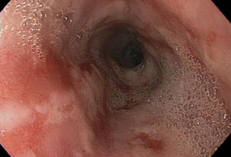
Patients with persistent symptoms on therapy with PPIs
Further testing is warranted in patients with persistent symptoms despite PPI therapy. Endoscopy should be performed, if not performed previously.[1]Katz PO, Dunbar KB, Schnoll-Sussman FH, et al. ACG clinical guideline for the diagnosis and management of gastroesophageal reflux disease. Am J Gastroenterol. 2022 Jan 1;117(1):27-56.
https://journals.lww.com/ajg/fulltext/2022/01000/acg_clinical_guideline_for_the_diagnosis_and.14.aspx
http://www.ncbi.nlm.nih.gov/pubmed/34807007?tool=bestpractice.com
[36]Yadlapati R, Gyawali CP, Pandolfino JE, et al. AGA clinical practice update on the personalized approach to the evaluation and management of GERD: expert review. Clin Gastroenterol Hepatol. 2022 May;20(5):984-94.e1.
https://www.cghjournal.org/article/S1542-3565(22)00079-9/fulltext
http://www.ncbi.nlm.nih.gov/pubmed/35123084?tool=bestpractice.com
In the absence of erosive esophagitis (Los Angeles grade B and above) or long-segment Barrett esophagus (≥3 cm), prolonged ambulatory pH monitoring should be performed, off drug therapy, to confirm or rule out GERD.[36]Yadlapati R, Gyawali CP, Pandolfino JE, et al. AGA clinical practice update on the personalized approach to the evaluation and management of GERD: expert review. Clin Gastroenterol Hepatol. 2022 May;20(5):984-94.e1.
https://www.cghjournal.org/article/S1542-3565(22)00079-9/fulltext
http://www.ncbi.nlm.nih.gov/pubmed/35123084?tool=bestpractice.com
Esophageal manometry should be performed:[1]Katz PO, Dunbar KB, Schnoll-Sussman FH, et al. ACG clinical guideline for the diagnosis and management of gastroesophageal reflux disease. Am J Gastroenterol. 2022 Jan 1;117(1):27-56.
https://journals.lww.com/ajg/fulltext/2022/01000/acg_clinical_guideline_for_the_diagnosis_and.14.aspx
http://www.ncbi.nlm.nih.gov/pubmed/34807007?tool=bestpractice.com
before antireflux surgery;
in patients unresponsive to PPIs where an etiology cannot be determined using impedance-pH monitoring;
and in patients with non-cardiac chest pain, especially those unresponsive to PPIs, to assess for motility abnormalities.
Other imaging modalities
Esophageal capsule endoscopy is a less-invasive, safe alternative to upper endoscopy, and a potential screening and diagnostic tool to evaluate esophageal pathology. Studies have shown only moderate sensitivity and specificity for diagnosis of esophageal disorders, and it has a limited role and acceptance in screening for mucosal disease (erosive esophagitis and Barrett esophagus).[38]Sharma P, Wani S, Rastogi A, et al. The diagnostic accuracy of esophageal capsule endoscopy: a blinded, prospective study. Am J Gastroenterol. 2008 Mar;103(3):525-32.
http://www.ncbi.nlm.nih.gov/pubmed/17459025?tool=bestpractice.com
[39]Eliakim R, Sharma VK, Yassin K, et al. A prospective study of the diagnostic accuracy of PillCam ESO esophageal capsule endoscopy versus conventional upper endoscopy in patients with chronic gastroesophageal reflux diseases. J Clin Gastroenterol. 2005 Aug;39(7):572-8.
http://www.ncbi.nlm.nih.gov/pubmed/16000923?tool=bestpractice.com
[40]Bhardwaj A, Hollenbeak CS, Pooran N. A meta-analysis of the diagnostic accuracy of esophageal capsule endoscopy. Am J Gastroenterol. 2009 Jun;104(6):1533-9.
http://www.ncbi.nlm.nih.gov/pubmed/19491867?tool=bestpractice.com
Capsule endoscopy is done for patient convenience in select circumstances. It is contraindicated in the presence of suspected (e.g., presence of dysphagia) or known stricture or adhesions.
Barium swallow may be useful in patients with dysphagia for whom endoscopy is contraindicated or unavailable.[41]Hirano I, Richter JE; Practice Parameters Committee of the American College of Gastroenterology. ACG Practice Guidelines: esophageal reflux testing. Am J Gastroenterol. 2007 Mar;102(3):668-85.
http://www.gi.org/physicians/guidelines/EsophagealRefluxTesting.pdf
http://www.ncbi.nlm.nih.gov/pubmed/17335450?tool=bestpractice.com
[42]Levine MS, Carucci LR, DiSantis DJ, et al. Consensus statement of Society of Abdominal Radiology disease-focused panel on barium esophagography in gastroesophageal reflux disease. AJR Am J Roentgenol. 2016 Nov;207(5):1009-15.
https://www.ajronline.org/doi/pdf/10.2214/AJR.16.16323
http://www.ncbi.nlm.nih.gov/pubmed/27490234?tool=bestpractice.com
Barium imaging should not be used solely as a diagnostic test for GERD.[1]Katz PO, Dunbar KB, Schnoll-Sussman FH, et al. ACG clinical guideline for the diagnosis and management of gastroesophageal reflux disease. Am J Gastroenterol. 2022 Jan 1;117(1):27-56.
https://journals.lww.com/ajg/fulltext/2022/01000/acg_clinical_guideline_for_the_diagnosis_and.14.aspx
http://www.ncbi.nlm.nih.gov/pubmed/34807007?tool=bestpractice.com
The presence of reflux on a barium esophagram has poor sensitivity and specificity for GERD, compared with pH testing.[1]Katz PO, Dunbar KB, Schnoll-Sussman FH, et al. ACG clinical guideline for the diagnosis and management of gastroesophageal reflux disease. Am J Gastroenterol. 2022 Jan 1;117(1):27-56.
https://journals.lww.com/ajg/fulltext/2022/01000/acg_clinical_guideline_for_the_diagnosis_and.14.aspx
http://www.ncbi.nlm.nih.gov/pubmed/34807007?tool=bestpractice.com