Images and videos
Images
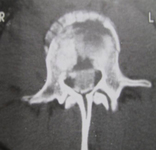

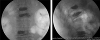

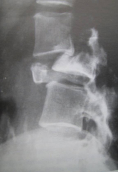
Osteoporotic spinal compression fractures
Anteroposterior and lateral x-ray images of patient with osteoporotic spinal compression fractures of L1,2,4 following kyphoplasty
Personal collection of Nasir A. Quraishi
See this image in context in the following section/s:
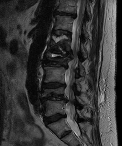
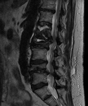
Osteoporotic spinal compression fractures
Preoperative sagittal T2-weighted magnetic resonance imaging showing osteoporotic spinal compression fractures of L1,2,4
Personal collection of Nasir A. Quraishi
See this image in context in the following section/s:
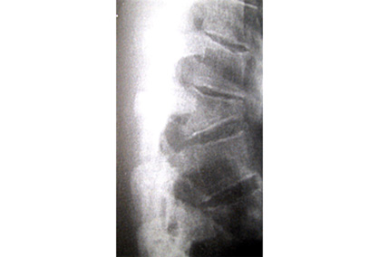
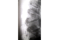
Osteoporotic spinal compression fractures
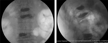
Lateral radiograph showing a T12 compression fracture in osteoporotic bone
Personal collection of Nasir A. Quraishi
See this image in context in the following section/s:
Use of this content is subject to our disclaimer