The possibility of a spinal compression fracture should be considered in patients at risk of osteoporotic bone disease, particularly older people and those taking long-term corticosteroid therapy.[50]American College of Radiology. ACR appropriateness criteria: thoracic back pain. 2024 [internet publication].
https://acsearch.acr.org/docs/3195158/Narrative
In the setting of “red flags” (e.g., history of significant trauma, minor fall or heavy lift in a potentially osteoporotic or elderly person, prolonged use of corticosteroids), the initial evaluation of a painful vertebral compression fracture includes assessing any neurologic deficits and evaluating mechanical versus radicular pain.[9]American College of Radiology. ACR Appropriateness Criteria: management of vertebral compression fractures. 2022 [internet publication].
https://acsearch.acr.org/docs/70545/Narrative
Presentation
Most osteoporotic spinal compression fractures are identified as an incidental finding on chest and abdominal x-rays taken for other indications, and these patients are frequently asymptomatic at the time of diagnosis.[11]LeBoff MS, Greenspan SL, Insogna KL, et al. The clinician's guide to prevention and treatment of osteoporosis. Osteoporos Int. 2022 Oct;33(10):2049-102.
https://link.springer.com/article/10.1007/s00198-021-05900-y
http://www.ncbi.nlm.nih.gov/pubmed/35478046?tool=bestpractice.com
[51]Alsoof D, Anderson G, McDonald CL, et al. Diagnosis and management of vertebral compression fracture. Am J Med. 2022 Jul;135(7):815-21.
http://www.ncbi.nlm.nih.gov/pubmed/35307360?tool=bestpractice.com
However, some patients may report an acute onset of back pain with relatively atraumatic activities, such as standing from a seated position, picking up a suitcase, bending forward, or even coughing or sneezing.[51]Alsoof D, Anderson G, McDonald CL, et al. Diagnosis and management of vertebral compression fracture. Am J Med. 2022 Jul;135(7):815-21.
http://www.ncbi.nlm.nih.gov/pubmed/35307360?tool=bestpractice.com
Patients may variably describe the pain as dull or sharp, and often as being aggravated by movement. Patients may report disturbance of sleep. The pain may radiate bilaterally to the abdomen, although, unlike pain from a herniated intervertebral disk, it rarely radiates to the legs. Local pain originating from vertebral fractures may last for 3 years or more; but many vertebral fractures do not cause any pain.[52]Johansson L, Svensson HK, Karlsson J, et al. Decreased physical health-related quality of life-a persisting state for older women with clinical vertebral fracture. Osteoporos Int. 2019 Oct;30(10):1961-71.
https://link.springer.com/article/10.1007/s00198-019-05044-0
http://www.ncbi.nlm.nih.gov/pubmed/31227884?tool=bestpractice.com
[53]Ross PD. Clinical consequences of vertebral fractures. Am J Med. 1997 Aug 18;103(2a):30S-42S; discussion 42S-3S.
http://www.ncbi.nlm.nih.gov/pubmed/9302895?tool=bestpractice.com
[54]Fechtenbaum J, Cropet C, Kolta S, et al. The severity of vertebral fractures and health-related quality of life in osteoporotic postmenopausal women. Osteoporos Int. 2005 Dec;16(12):2175-9.
http://www.ncbi.nlm.nih.gov/pubmed/16220230?tool=bestpractice.com
Patients with multiple fractures and a marked kyphosis may report weight gain and difficulty fitting into clothes, although their weight remains stable. This is due to loss of height, and the kyphosis compressing abdominal contents and causing the abdomen to bulge forward. Progressive kyphosis of the thoracic spine with compensatory lumbar lordosis can result in decreased appetite.[51]Alsoof D, Anderson G, McDonald CL, et al. Diagnosis and management of vertebral compression fracture. Am J Med. 2022 Jul;135(7):815-21.
http://www.ncbi.nlm.nih.gov/pubmed/35307360?tool=bestpractice.com
Severe kyphosis or multiple vertebral fractures can affect pulmonary function and may lead to dyspnea.[11]LeBoff MS, Greenspan SL, Insogna KL, et al. The clinician's guide to prevention and treatment of osteoporosis. Osteoporos Int. 2022 Oct;33(10):2049-102.
https://link.springer.com/article/10.1007/s00198-021-05900-y
http://www.ncbi.nlm.nih.gov/pubmed/35478046?tool=bestpractice.com
[55]Harrison RA, Siminoski K, Vethanayagam D, et al. Osteoporosis-related kyphosis and impairments in pulmonary function: a systematic review. J Bone Miner Res. 2007 Mar;22(3):447-57.
https://asbmr.onlinelibrary.wiley.com/doi/full/10.1359/jbmr.061202
http://www.ncbi.nlm.nih.gov/pubmed/17181402?tool=bestpractice.com
These patients may also report neck pain resulting from the need for continuous neck extension to look forward or upward.
Lumbar fractures can alter abdominal anatomy, leading to constipation, abdominal pain, early satiety, and weight loss.[11]LeBoff MS, Greenspan SL, Insogna KL, et al. The clinician's guide to prevention and treatment of osteoporosis. Osteoporos Int. 2022 Oct;33(10):2049-102.
https://link.springer.com/article/10.1007/s00198-021-05900-y
http://www.ncbi.nlm.nih.gov/pubmed/35478046?tool=bestpractice.com
Loss of sagittal balance occurs when the patient is no longer able to compensate for progressive kyphosis by rotating the pelvis backward.[56]Fechtenbaum J, Etcheto A, Kolta S, et al. Sagittal balance of the spine in patients with osteoporotic vertebral fractures. Osteoporos Int. 2016 Feb;27(2):559-67.
http://www.ncbi.nlm.nih.gov/pubmed/26272312?tool=bestpractice.com
This causes intolerance of standing still or walking slowly. Sometimes this can be compensated for by walking fast, or by using impromptu walking aids, such as a grocery cart or child's stroller.
Clinical exam
Patients with multiple wedge fractures of the thoracic spine may have a notable kyphosis, although this can also occur in the absence of vertebral fractures. Conversely, wedge fractures of the lumbar spine can lead to lessening of the lumbar lordosis. Loss of height also occurs with these fractures, particularly if there are multiple fractures.[1]Kim DH, Vaccaro AR. Osteoporotic compression fractures of the spine: current options and considerations for treatment. Spine J. 2006 Sep-Oct;6(5):479-87.
http://www.ncbi.nlm.nih.gov/pubmed/16934715?tool=bestpractice.com
[51]Alsoof D, Anderson G, McDonald CL, et al. Diagnosis and management of vertebral compression fracture. Am J Med. 2022 Jul;135(7):815-21.
http://www.ncbi.nlm.nih.gov/pubmed/35307360?tool=bestpractice.com
In combination with a kyphosis, this loss of height can result in bulging of the abdomen as the contents are compressed and pushed forward. As a consequence of these factors, the patient may not be able to stand upright with the head balanced over the hips without bending the knees (loss of sagittal balance).[56]Fechtenbaum J, Etcheto A, Kolta S, et al. Sagittal balance of the spine in patients with osteoporotic vertebral fractures. Osteoporos Int. 2016 Feb;27(2):559-67.
http://www.ncbi.nlm.nih.gov/pubmed/26272312?tool=bestpractice.com
In an acute injury, there will be tenderness locally over the spine at the level involved. A full neurologic exam is indicated in these patients. Neurologic exam is also indicated after a minor fall or heavy lift in a potentially osteoporotic or elderly person or in patients with a history of prolonged use of corticosteroids.[9]American College of Radiology. ACR Appropriateness Criteria: management of vertebral compression fractures. 2022 [internet publication].
https://acsearch.acr.org/docs/70545/Narrative
Although osteoporotic spinal compression fractures do not generally cause neurologic problems, the presence of neurologic signs indicates a need for urgent computed tomography (CT) or magnetic resonance imaging (MRI).
Diagnostic workup
Where there are concerns about the occurrence of a vertebral fracture(s), recent imaging that includes the spine should be reviewed. If no recent imaging is available or the patient’s symptoms began after the previous imaging, patients should be referred for spine imaging. The referral should highlight the concern about the presence of fracture.[57]Royal Osteoporosis Society. Guidance for the management of symptomatic vertebral fragility fractures. May 2022 [internet publication].
https://strwebprdmedia.blob.core.windows.net/media/kuphgv1u/ros-guidance-on-managing-symptoms-of-vertebral-fractures-2022.pdf
Plain x-ray
Initial investigation for all patients involves radiographic evaluation with anteroposterior (AP) and lateral spine x-rays.[11]LeBoff MS, Greenspan SL, Insogna KL, et al. The clinician's guide to prevention and treatment of osteoporosis. Osteoporos Int. 2022 Oct;33(10):2049-102.
https://link.springer.com/article/10.1007/s00198-021-05900-y
http://www.ncbi.nlm.nih.gov/pubmed/35478046?tool=bestpractice.com
These often reveal the classic wedge fracture with loss of anterior vertebral height and relative preservation of posterior body height. In osteoporotic patients, fractures usually occur around the midthoracic level (T7-T8) or thoracolumbar junction level (T12-L1). The AP x-ray may show interpedicular widening or malalignment of the spinous processes, which would suggest posterior column injury. Other radiographic features of concern include loss of vertebral height >50% or segmental kyphosis >20° (both suggest posterior ligamentous injury), or multiple adjacent compression fractures that may require surgical treatment to reduce the development of kyphotic deformity.
Distinguishing acute fractures from old fractures on plain radiographs is difficult.[1]Kim DH, Vaccaro AR. Osteoporotic compression fractures of the spine: current options and considerations for treatment. Spine J. 2006 Sep-Oct;6(5):479-87.
http://www.ncbi.nlm.nih.gov/pubmed/16934715?tool=bestpractice.com
Acute fractures typically have well-demarcated fracture lines or distinct discontinuity of the thin layer of cortical bone. Older fractures often have sclerosis of the fracture lines, a dense cortical margin, and osteophyte formation around the fracture site. In cases where there are no previous plain radiographs for comparison, MRI or bone scan may be needed to age fracture acuity.[50]American College of Radiology. ACR appropriateness criteria: thoracic back pain. 2024 [internet publication].
https://acsearch.acr.org/docs/3195158/Narrative
[Figure caption and citation for the preceding image starts]: Lateral radiograph showing a T12 compression fracture in osteoporotic bonePersonal collection of Nasir A. Quraishi [Citation ends].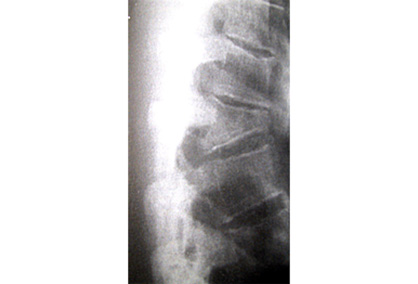
MRI spine/CT spine
In patients with new symptomatic vertebral compression fractures identified on radiographs and no known malignancy, MRI or CT spine of the area of interest is usually appropriate for pre-procedural planning, if intervention is being considered.[9]American College of Radiology. ACR Appropriateness Criteria: management of vertebral compression fractures. 2022 [internet publication].
https://acsearch.acr.org/docs/70545/Narrative
[50]American College of Radiology. ACR appropriateness criteria: thoracic back pain. 2024 [internet publication].
https://acsearch.acr.org/docs/3195158/Narrative
CT or MRI spine of the area of interest are particularly useful for initial imaging if radiographs are negative.[50]American College of Radiology. ACR appropriateness criteria: thoracic back pain. 2024 [internet publication].
https://acsearch.acr.org/docs/3195158/Narrative
CT provides osseous details of axial spine fractures before vertebral augmentation and permits evaluation of vertebral body height, architecture, and integrity of the posterior cortex and pedicles before vertebral augmentation, which is critical in patients with cortical disruption, posterior cortex osseous retropulsion, and spinal canal compression.[9]American College of Radiology. ACR Appropriateness Criteria: management of vertebral compression fractures. 2022 [internet publication].
https://acsearch.acr.org/docs/70545/Narrative
MRI is particularly useful in identifying minimally compressed fractures, distinguishing acute from chronic fractures, and distinguishing fractures due to tumor or infection.[1]Kim DH, Vaccaro AR. Osteoporotic compression fractures of the spine: current options and considerations for treatment. Spine J. 2006 Sep-Oct;6(5):479-87.
http://www.ncbi.nlm.nih.gov/pubmed/16934715?tool=bestpractice.com
[9]American College of Radiology. ACR Appropriateness Criteria: management of vertebral compression fractures. 2022 [internet publication].
https://acsearch.acr.org/docs/70545/Narrative
[50]American College of Radiology. ACR appropriateness criteria: thoracic back pain. 2024 [internet publication].
https://acsearch.acr.org/docs/3195158/Narrative
If there are neurologic signs, concerns over fracture stability, or a suspicion that fractures may be pathologic, a CT scan and/or an MRI is indicated.[19]American College of Surgeons. Best practices guidelines. Spine injury. Mar 2022 [internet publication].
https://www.facs.org/media/k45gikqv/spine_injury_guidelines.pdf
MRI is more useful than CT for assessing the integrity of the soft tissue and neural structures, particularly that of the spinal canal.[50]American College of Radiology. ACR appropriateness criteria: thoracic back pain. 2024 [internet publication].
https://acsearch.acr.org/docs/3195158/Narrative
MRI is the only modality for evaluating the internal structure of the spinal cord.[58]American College of Radiology. ACR–ASNR–SCBT-MR–SSR practice parameter for the performance of magnetic resonance imaging (MRI) of the adult spine. 2018 [internet publication].
https://www.acr.org/-/media/ACR/Files/Practice-Parameters/mr-adult-spine.pdf
Technetium-99m (Tc-99m) whole-body bone scan (bone scintigraphy)
Tc-99m whole-body bone scan (bone scintigraphy) may be helpful to determine the painful vertebrae, particularly the causative level.[9]American College of Radiology. ACR Appropriateness Criteria: management of vertebral compression fractures. 2022 [internet publication].
https://acsearch.acr.org/docs/70545/Narrative
Whole-body bone scans may be helpful in the setting of compression fractures to help identify fracture acuity and to appropriately select patients for intervention, particularly if MRI cannot be safely/easily obtained.[50]American College of Radiology. ACR appropriateness criteria: thoracic back pain. 2024 [internet publication].
https://acsearch.acr.org/docs/3195158/Narrative
[59]Ramachandran S, Clifton IJ, Collyns TA, et al. The treatment of spinal tuberculosis: a retrospective study. Int J Tuberc Lung Dis. 2005 May;9(5):541-4.
http://www.ncbi.nlm.nih.gov/pubmed/15875926?tool=bestpractice.com
Single-photon emission computed tomography (SPECT)/CT
SPECT coupled with CT may also be appropriate.[9]American College of Radiology. ACR Appropriateness Criteria: management of vertebral compression fractures. 2022 [internet publication].
https://acsearch.acr.org/docs/70545/Narrative
[50]American College of Radiology. ACR appropriateness criteria: thoracic back pain. 2024 [internet publication].
https://acsearch.acr.org/docs/3195158/Narrative
SPECT/CT has been shown to localize abnormalities in the vertebra more precisely compared with SPECT imaging alone, particularly in complicated cases, such as multiple collapsed vertebrae of different ages.[9]American College of Radiology. ACR Appropriateness Criteria: management of vertebral compression fractures. 2022 [internet publication].
https://acsearch.acr.org/docs/70545/Narrative
[60]Kumar K, Halkar RK, Bartley SC, et al. Incremental benefit of SPECT + CT bone scans over conventional planar and SPECT bone scans in vertebroplasty. Indian J Nucl Med. 2011 Oct;26(4):181-4.
https://www.ncbi.nlm.nih.gov/pmc/articles/PMC3613623
http://www.ncbi.nlm.nih.gov/pubmed/23559712?tool=bestpractice.com
SPECT/CT appears to be comparable to MRI in detecting fractures, particularly in the acute phase, and could be considered if MRI is contraindicated.[61]Li YB, Zheng X, Wang R, et al. SPECT-CT versus MRI in localizing active lesions in patients with osteoporotic vertebral compression fractures. Nucl Med Commun. 2018 Jul;39(7):610-7.
http://www.ncbi.nlm.nih.gov/pubmed/29893749?tool=bestpractice.com
[62]Lems WF, Paccou J, Zhang J, et al. Vertebral fracture: epidemiology, impact and use of DXA vertebral fracture assessment in fracture liaison services. Osteoporos Int. 2021 Mar;32(3):399-411.
https://link.springer.com/article/10.1007/s00198-020-05804-3
http://www.ncbi.nlm.nih.gov/pubmed/33475820?tool=bestpractice.com
Other investigations
Additional investigations may be appropriate:
If a diagnosis of osteoporosis is suspected but unconfirmed, a nonurgent dual-energy x-ray absorptiometry bone density scan may be obtained as part of the workup.[11]LeBoff MS, Greenspan SL, Insogna KL, et al. The clinician's guide to prevention and treatment of osteoporosis. Osteoporos Int. 2022 Oct;33(10):2049-102.
https://link.springer.com/article/10.1007/s00198-021-05900-y
http://www.ncbi.nlm.nih.gov/pubmed/35478046?tool=bestpractice.com
If there is a suspicion of malignancy, a complete blood count (CBC), serum alkaline phosphatase, and C-reactive protein are indicated.
If there is a suspicion of infection, a CBC and blood cultures should be obtained.
A bone profile may be useful to exclude a metabolic cause. This includes serum calcium, albumin, parathyroid hormone, phosphate, alkaline phosphatase, magnesium, creatinine, and serum 25-hydroxyvitamin D (25OHD).
Other tests such as thyroid-stimulating hormone, screening for hypercortisolism, and serum protein electrophoresis may also be considered.
If spinal hardware is present, CT myelography may be useful in the assessment of hardware integrity and position.[50]American College of Radiology. ACR appropriateness criteria: thoracic back pain. 2024 [internet publication].
https://acsearch.acr.org/docs/3195158/Narrative
[63]Patel DM, Weinberg BD, Hoch MJ. CT myelography: clinical indications and imaging findings. Radiographics. 2020 Mar-Apr;40(2):470-84.
https://pubs.rsna.org/doi/10.1148/rg.2020190135
http://www.ncbi.nlm.nih.gov/pubmed/32058837?tool=bestpractice.com