Images and videos
Images

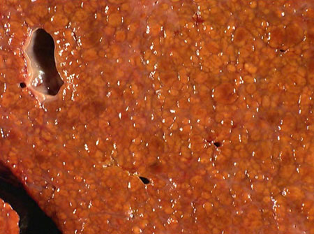
Steatotic liver disease
A native hepatectomy specimen from a patient with a history of metabolic dysfunction-associated steatohepatitis; a cross-section of the liver shows mixed, predominantly micronodular cirrhosis; the orange-yellow color of the nodules is due to moderate mixed micro- and macrovesicular steatosis involving 40% of the liver parenchyma
From the collection of Kapil B. Chopra, MD
See this image in context in the following section/s:
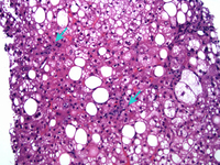
Steatotic liver disease
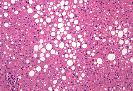
A case of metabolic dysfunction-associated steatohepatitis; the biopsy shows ballooning degeneration of the hepatocytes (middle right) and spotty lobular inflammation in addition to mixed micro- and macrovesicular steatosis (H&E, x 200)
From the collection of Kapil B. Chopra, MD
See this image in context in the following section/s:
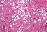
Steatotic liver disease
A wedge biopsy of the liver from a 52-year-old female organ donor; the biopsy shows moderate mixed micro- and macrovesicular steatosis; there is no significant lobular inflammation or necrosis (hematoxylin and eosin, [H&E] stain, x 200)
From the collection of Kapil B. Chopra, MD
See this image in context in the following section/s:
Use of this content is subject to our disclaimer