Images and videos
Images


Evaluation of pustular rash
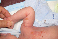
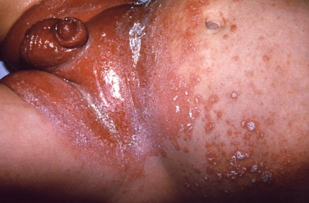
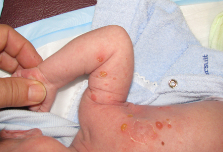
Infant presenting with rash formerly known as moniliasis, now called candidiasis, caused by Candida spp
Public Health Image Library, CDC
See this image in context in the following section/s:

Evaluation of pustular rash
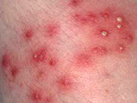
Superficial folliculitis with prominent erythematous papules and pustules
From the collection of Dr Professor Baden; used with permission
See this image in context in the following section/s:
Evaluation of pustular rash
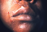
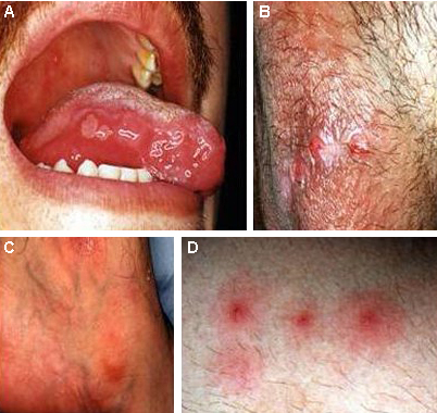
Mouth and genital ulcers in Behcet disease
From the collection of Dr Yusuf Yazici; used with permission
See this image in context in the following section/s:
Evaluation of pustular rash
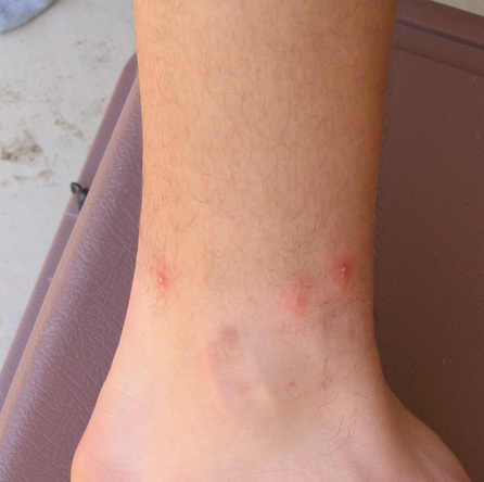
Pseudopustule formation following fire ant sting
From the collection of Theodore Freeman; used with permission
See this image in context in the following section/s:

Evaluation of pustular rash
Pustules in a patient with reactive arthritis
Public Health Image Library, CDC
See this image in context in the following section/s:
Evaluation of pustular rash
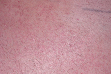
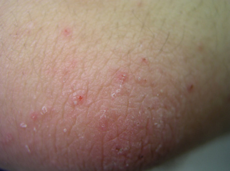
Miliaria rubra
From the collection of Brian L. Swick; used with permission
See this image in context in the following section/s:
Evaluation of pustular rash
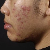
Nodulocystic acne
University of Michigan Department of Dermatology
See this image in context in the following section/s:

Evaluation of pustular rash
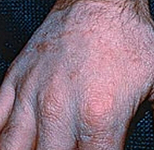
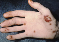
Tinea manuum. On the extensor surface of the hand there is extensive inflammation, scaling, hyperkeratosis, and erythema
Department of Dermatology Medical University of South Carolina; used with permission
See this image in context in the following section/s:
Evaluation of pustular rash
Neonate with bullous impetigo
From the collection of Michael Freeman; used with permission
See this image in context in the following section/s:
Evaluation of pustular rash
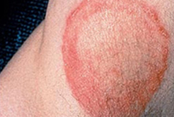
Tinea corporis of the axilla. Central clearing with an active border of inflammation noted. Satellite lesion is present
Department of Dermatology Medical University of South Carolina; used with permission
See this image in context in the following section/s:

Evaluation of pustular rash
Secondary syphilitic lesions on the face
Public Health Image Library, CDC
See this image in context in the following section/s:

Evaluation of pustular rash
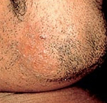
Tinea barbae. Note the pustules in the follicles, redness, and scaling
Department of Dermatology Medical University of South Carolina; used with permission
See this image in context in the following section/s:
Evaluation of pustular rash
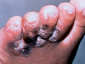
Tinea pedis. Intense inflammation produces hyperpigmentation and vesicle formation. Vesiculobullous form of tinea pedis
Department of Dermatology Medical University of South Carolina; used with permission
See this image in context in the following section/s:

Evaluation of pustular rash
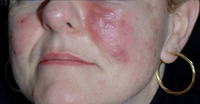
Rosacea fulminans
Courtesy of Dr Richard Allen Johnson (MD, CM. Harvard Medical School); used with permission
See this image in context in the following section/s:
Evaluation of pustular rash
Scabies: characteristic linear burrows in skin
From the collection of Dr Laura Ferris; used with permission
See this image in context in the following section/s:
Use of this content is subject to our disclaimer