Tests
Tests to consider
anteroposterior and lateral radiographs of foot
Test
Not routinely ordered in classic clubfoot deformities.[21] Lateral view should be in maximum dorsiflexion. Kite angle is measured by drawing a line down the center of the calcaneus and talus. This is about 5° in clubfeet, with a greater angle in normal and isolated metatarsus adductus.[30] Should be ordered if a limb deficiency (e.g., fibular hemimelia, tibial hemimelia, or congenital short femur) is noted on examination.[21]
Result
parallelism between talus and calcaneus
dynamic hip ultrasound
Test
Helps establish diagnosis of hip dysplasia in infants. May be false-positive if done too early. Routinely performed at many centers despite uncertainty regarding the association between hip dysplasia and clubfeet.[23][24][25] The British Society for Children’s Orthopaedic Surgery (BSCOS) Clubfoot Consensus Group has recommended that all babies with a clubfoot deformity should receive a screening ultrasound scan of their hips.[22]
Result
hip dysplasia: subluxation on provocative testing; abnormal relationship between femoral head and acetabulum
pelvic radiographs
Test
Helps establish a diagnosis of hip dysplasia. The ossific nucleus of the proximal femur is not usually ossified until after age 4 months.
Routinely performed at many centers despite uncertainty regarding the association between hip dysplasia and clubfeet.[23][24][25]
Result
abnormal relationship between femoral head and acetabulum (assessed by acetabular index, Shenton line, ossification of femoral head)
CT foot
Test
Performed in older children or adolescents to help with preoperative planning. [Figure caption and citation for the preceding image starts]: Three-dimensional view of clubfoot deformity in an adolescent, showing the classic equinus, varus, and adduction deformitiesFrom the collection of Scott E. Van Valin [Citation ends].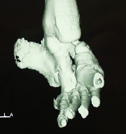
Result
deformity
CT spine
Test
Any evidence of signs such as dimpling or hairy patches of the spine on physical exam may suggest an underlying spinal abnormality and warrants further investigation.
Result
coexisting spinal abnormalities
abdominal ultrasound
Test
Any evidence of signs such as dimpling or hairy patches of the spine on physical exam may suggest an underlying spinal abnormality and warrants further investigation.
Result
coexisting spinal abnormalities
Use of this content is subject to our disclaimer