Approach
Viral infections are the most common cause of labyrinthitis. They typically occur in adults, whereas purulent bacterial labyrinthitis is more common in children who are otitis prone. Serous, as opposed to suppurative, labyrinthitis is still far more common in both pediatric and adult patients.
The differential diagnosis of patients presenting with dizziness and vertigo can often be narrowed with a thorough history and physical exam. Imaging studies (e.g., magnetic resonance imaging [MRI] and computed tomography [CT]) are typically ordered as patients with posterior fossa neoplasms can have a variety of presentations that can mimic other vestibular disorders. Anyone presenting with unilateral inner ear type symptoms should be investigated for retrocochlear pathology. Posterior fossa tumors include vestibular schwannomas (acoustic neuroma), meningiomas, cerebellar or brainstem tumors, and epidermoid cysts.
History
Patients with labyrinthitis typically present with severe room-spinning vertigo and associated nausea and vomiting. They may have unilateral hearing loss and tinnitus (ringing in the ear). The hearing loss is sensorineural (i.e., related to inner ear/eighth cranial nerve) rather than conductive (i.e., secondary to middle ear causes). Significant hearing loss, with or without associated tinnitus, differentiates labyrinthitis from vestibular neuritis.[1] Acute vertigo may last up to 72 hours and is typically followed by persistent disequilibrium (problems with balance) and brief vertigo (lasting seconds) that is associated with quick head or body movements.[4] If the patient experiences more than one episode of room-spinning vertigo, a diagnosis of Ménière disease should be considered. Although vertigo may be caused by infectious conditions, there are a range of potential causes that need to be ruled out (e.g., post-traumatic vertigo, or vertigo of cerebrovascular origin).
Important questions that are critical in establishing the diagnosis include:
Can you describe your dizziness (e.g., room spinning, imbalance, floating, lightheadedness)? Dizziness is a broad term, and this question helps to determine the actual sensation experienced by the patient.
Tell me about your first episode of vertigo. How long did this episode last? This can help the clinician determine if the patient has had labyrinthitis or vestibular neuritis.
How long are your current dizzy spells? Examining the duration of the patient's dizzy spells can provide critical information for the diagnosis.
Have you ever had any prior episodes of dizziness or vertigo? Recurrent vertigo also helps to narrow down the differential diagnosis.
Do you have any other associated symptoms (i.e., hearing loss, tinnitus, ear fullness, etc.)? Auditory symptoms concurrent with dizziness make a peripheral source of the dizziness more likely.
Do changes in body position cause dizziness? A positive response to this question narrows the differential to the following: benign paroxysmal positional vertigo, orthostasis, uncompensated peripheral vestibulopathy, or bilateral vestibulopathy.
Do you have any history of headaches (e.g., migraine, muscle tension)? A positive response increases the likelihood that migraine may be the source of dizziness.
Have you started any new (or changed) any medications? There are a number of medications that can cause dizziness.
What makes your dizziness better or worse? Movement associated dizziness helps narrow the differential diagnosis.
Have you recently had an upper respiratory tract infection? A positive response may indicate vestibular neuritis.
It is critical to evaluate for other neurologic symptoms such as dysarthria, dysphagia, facial pain or numbness, facial weakness, and extremity weakness or numbness, as these would point to a cerebrovascular accident involving the brainstem.[15]
Several tools, such as the Dizziness Handicap Inventory and the SF-36, can be used to assess the impact of dizziness on patients’ quality of life.[16]
Another approach that focuses on assessing dizziness, known as the TiTrATE (Timing, Triggers, And Targeted Exams) approach, examines the timing and potential triggers for symptoms to help diagnose the cause.[17]
Physical exam
Patients presenting in the acute setting may have significant difficulty walking. Spontaneous horizontal-rotary nystagmus (rapid involuntary movement of the eyes) with the fast phase beating toward the uninvolved ear is also frequently present.[18]
The patient's balance should be assessed using tandem gait and Romberg testing.[18] Patients will probably not be able to perform tandem gait (walking with one foot directly in front of the other foot, heel-to-toe) and may fall with Romberg testing (standing straight up with feet together and eyes closed).
Examination with a Weber 512-Hz tuning fork (placing the tuning fork on the forehead or maxillary teeth and asking the patient to state in which ear the sound was louder) can quickly localize the affected ear and determine whether the hearing loss is sensorineural or conductive. The sound will be perceived in the affected ear when a unilateral conductive hearing loss is present or in the unaffected ear when there is a unilateral sensorineural hearing loss. The result of this test is combined with the result of the Rinne test to interpret the type of hearing loss.[19]
Rinne testing allows the examiner to determine whether any hearing loss is secondary to middle ear (conductive hearing loss) or inner ear/eighth cranial nerve (sensorineural hearing loss) causes. The base of a 512-Hz tuning fork is placed on the mastoid and the patient indicates when he or she no longer hears the sound. Once the sound is no longer audible, the tuning fork is placed in front of the ear and the patient is asked whether he or she hears the sound. If the sound is louder when the tuning fork is on the mastoid, then the patient has a conductive hearing loss. If the sound is louder with the fork in front of the ear, the hearing loss is sensorineural or normal.
A thorough examination of the ear with an otoscope or microscope allows diagnosis of otitis media and cholesteatoma. The presence of otorrhea (ear discharge) should alert the clinician to the presence of acute or chronic otitis media with tympanic membrane perforation. Careful inspection of the entire tympanic membrane should identify a cholesteatoma unless there is significant debris in the ear canal that obscures visualization.[Figure caption and citation for the preceding image starts]: Cholesteatoma in middle earFrom the collection of Professor Brandon Isaacson; used with permission [Citation ends].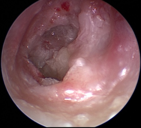
There should be no evidence of other neurologic deficits such as upper or lower extremity weakness, hoarseness, or facial weakness or numbness. The three-step bedside oculomotor exam Head-Impulse, Nystagmus, Test-of-Skew (HINTS) has been found to identify stroke with a high degree of sensitivity and specificity in patients with acute vestibular symptoms, and it may rule out stroke more effectively than early diffusion-weighted MRI.[20] Based on the HINTS model, one algorithm suggests that stroke should be considered in patients presenting with acute-onset dizziness if:[21]
there is a central pattern of nystagmus
there is skew deviation
there is a negative head impulse test (in patients with nystagmus)
there are any central nervous system signs on focused neurologic examination, or
the patient is unable to sit or walk unaided.
The presence of meningeal signs should be investigated if bacterial meningitis is a consideration. For example, a rash is noted in 80% to 90% of patients with meningococcal meningitis, most commonly 4 to 18 hours after the initial symptoms of illness. Typically, the rash is a nonblanching petechial or purpuric exanthem, but a few patients may initially have nonspecific erythematous macular or maculopapular lesions.
Cerebellar function should be examined by requesting the patient to perform finger to nose, heel to shin, and rapid alternating movement tests.[15]
Audiometry
An audiometric exam is useful to document the extent of hearing loss and to confirm the affected ear. Hearing loss is typically of the sensorineural type. However, patients with inner ear malformations (i.e., enlarged vestibular aqueduct) may present with similar symptoms and a mixed hearing loss with a significant sensorineural component.
Vestibular testing with electronystagmography, rotary chair test, and vestibular-evoked myogenic potentials is not indicated in the acute setting. However, these tests may provide additional information on vestibular compensation and site-of-lesion testing after the patient has recovered from the acute stage of labyrinthitis.[18]
Laboratory tests
Patients with labyrinthitis secondary to bacterial meningitis should have appropriate cerebrospinal fluid cultures performed. Additional serologic testing for syphilis and HIV may be warranted if the presentation is atypical or if the patient has additional risk factors.[22] If the serologic tests are negative, autoimmune conditions (e.g., Cogan syndrome or Behcet disease) may be suspected.[5] For patients who have severe nausea and vomiting, a basic metabolic panel should be evaluated to select the appropriate crystalloid and electrolyte replacement. Check capillary blood glucose in all patients with suspected stroke and arrange urgent neuroimaging.[23]
Imaging
Imaging can help to rule out differential diagnoses.
If an acute stroke is suspected, a CT scan of the head can identify infarction and provide enough information to make decisions about acute management.[23] A subsequent MRI of the head with diffusion-weighted imaging can determine the extent of the infarct. If a temporal bone fracture is suspected, a CT scan of the head can delineate the extent of the fracture.[24] MRI or CT scans of the head can reveal inner ear malformations and temporal bone neoplasms. A CT scan of the petrous temporal bones may show evidence of middle-ear or mastoid opacification, and should be ordered if the patient is suspected of having mastoiditis. A CT scan may also be useful in patients with suspected superior semicircular canal dehiscence, and also to confirm cholesteatoma.[25][26]
Any patient with an asymmetric hearing loss should undergo a retrocochlear evaluation with gadolinium-enhanced MRI to investigate other causes of hearing loss. For example, over 10% of patients with vestibular schwannomas (acoustic neuromas) present with sudden hearing loss. Labyrinthine enhancement on gadolinium-enhanced MRI in the setting of meningitis is a significant predictor of hearing loss.[27]
Use of this content is subject to our disclaimer