Tests
1st tests to order
pulse oximetry
Test
Preductal and postductal (right arm and either leg) pulse oximetry should be ordered if there is any suspicion of a congenital cardiac malformation.[19]
May be normal in TOF with mild pulmonary stenosis. However, in TOF with moderate to severe pulmonary stenosis, the baby is hypoxemic.
Result
low oxygen saturation
echocardiogram
Test
Should be ordered in any newborn with a suspected diagnosis of congenital heart disease. Echocardiography is the definitive investigation for diagnosis of TOF.
Result
infundibular pulmonic stenosis, overriding aorta, nonrestrictive ventricular septal defect, concentric right ventricular hypertrophy
ECG
Test
Right ventricular hypertrophy (RVH) may be difficult to interpret in a neonate. [Figure caption and citation for the preceding image starts]: ECG in tetralogy of Fallot showing right ventricular hypertrophyFrom the collection of Dr Jeffrey Gossett; used with permission [Citation ends].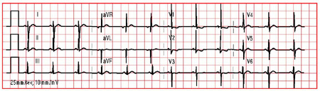
In an older child RVH is more likely to be seen.
Result
RVH with a rightward axis, R in V1 and S in V6 above age-appropriate normals
CXR
Test
Normal cardiac silhouette does not rule out cyanotic heart disease.
Result
boot-shaped heart
hyperoxygenation test
Test
Used to determine whether hypoxemia is from a pulmonary or a cardiac lesion.
PaO2 on room air should be checked, 100% FiO2 given for at least 10 minutes, and then PaO2 rechecked. If the PaO2 increases by >25 mmHg and to >100 mmHg, the hypoxemia is likely to be caused by a pulmonary problem.
In acyanotic patients this test may yield false-negative results.
Result
no significant increase in PaO2
Tests to consider
cardiac CT angiography or MRI
Test
Not routinely needed for evaluation in infants. Can be considered if definition of the coronary artery anatomy is not possible by echocardiography.[20]
Result
full definition of coronary anatomy
cardiac catheterization
Test
Not routinely done for diagnostic evaluation of TOF because stimulation of the infundibular muscle may precipitate hypercyanotic spells.
Occasionally intervention into the right ventricular outflow tract (RVOT) may be undertaken prior to complete repair with options including RVOT ballooning or stenting.[22]
Result
pulmonary stenosis and ventricular septal defects may be seen on angiogram; provides hemodynamic data such as systemic right ventricular pressure and right-to-left shunt
Use of this content is subject to our disclaimer