History and exam
Key diagnostic factors
common
asymptomatic grouped annular pink or flesh-colored dermal papules
Typical of localized disease.[Figure caption and citation for the preceding image starts]: Classic localized granuloma annulareFrom the collection of Susmita Mukherjee, BSc, MBBS, MRCP; used with permission [Citation ends].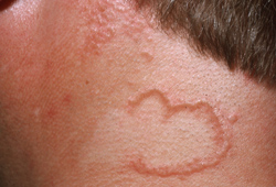
Papules arranged as a ring-like lesion.
Commonly found on the fingers in children and the shins of middle-aged women, although can occur anywhere on the body.
Other diagnostic factors
common
flesh-colored, pink, or brown macules or small papules
Typical of generalized disease.[Figure caption and citation for the preceding image starts]: Generalized granuloma annulareFrom the collection of Susmita Mukherjee, BSc, MBBS, MRCP; used with permission [Citation ends].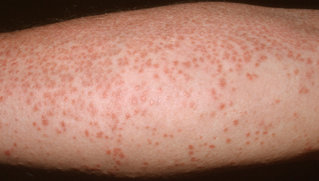 [Figure caption and citation for the preceding image starts]: Generalized granuloma annulareFrom the collection of Susmita Mukherjee, BSc, MBBS, MRCP; used with permission [Citation ends].
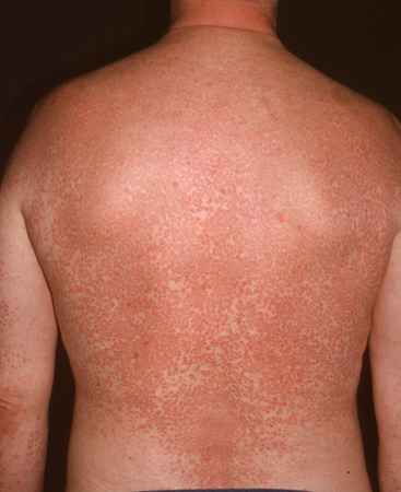
May number many thousands; predominantly found over chest in a symmetric pattern.
uncommon
soft-tissue nodules
Typical of subcutaneous disease.
Firm subcutaneous nontender lump without overlying skin changes.
perforating papules, crusting or ulcerated lesions
Typical of perforating disease.
Can be single or multiple.
More common in older patients.
erythematous patches
Typical of patch disease.
Very rare.
Palpable border with scattered papules.
Risk factors
weak
diabetes mellitus
hematologic malignancy
Evidence from case series suggests a potential link between GA and mycosis fungoides.[16]
Reactive T-cells in patients with Hodgkin lymphoma may cause GA by release of cytokines and metalloproteinases.[6]
Cases of GA in association with non-Hodgkin lymphoma, chronic myelomonocytic leukemia, and myelodysplastic syndrome have also been reported.[8][15][23]
Conversely, a cohort study of 60 patients with generalized GA compared to 300 controls identified a similar rate of malignancies in both populations, suggesting no increased risk of malignancy in patients with GA.[24] Age appropriate screening is recommended.[25]
herpes zoster
A case series described the occurrence of localized and perforating GA within herpes zoster scars, suggesting a potential causative role for herpes zoster.[26]
HIV
There is an association described between HIV and GA (termed HIV-associated GA). This may be generalized or localized to atypical sites such as the penis.[27] The exact mechanism is unknown. Perforating GA may be more common among people with HIV.
hepatitis
hyperlipidemia
One case-control study demonstrated increased incidence of dyslipidemia in patients with GA compared with controls.[14]
thyroid disease
medications
Drug-induced GA has been reported, most often with TNF-alpha inhibitors, although cases have also been reported with calcitonin, amlodipine, allopurinol, diclofenac, and intramuscular gold.[17] Some reports describe granulomatous reactions, including GA and GA-like eruptions, with targeted chemotherapeutic agents and immunotherapy drugs.[18][19]
Cases of GA developing after bacilli Calmette-Guerin (BCG) vaccination have also been reported.[30][31]
Use of this content is subject to our disclaimer