Images and videos
Images


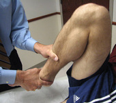


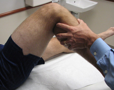
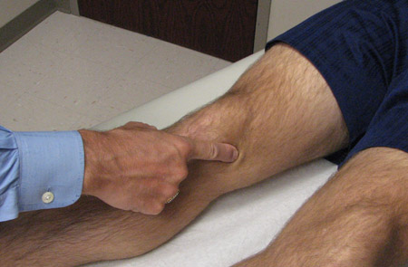
Evaluation of knee injury
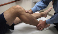
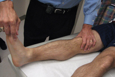
Posterolateral drawer test
From the personal library of Dr LaPrade; used with permission
See this image in context in the following section/s:
Evaluation of knee injury
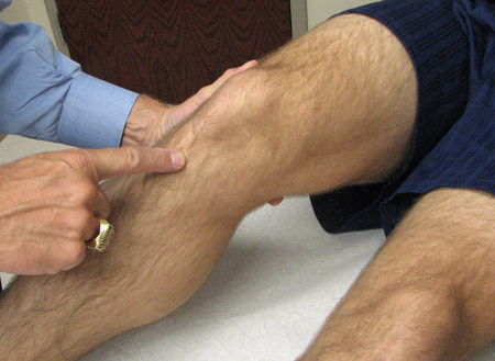
Figure 4 test
From the personal library of Dr LaPrade; used with permission
See this image in context in the following section/s:
Evaluation of knee injury
Pivot shift test
From the personal library of Dr LaPrade; used with permission
See this image in context in the following section/s:

Evaluation of knee injury
Valgus stress test
From the personal library of Dr LaPrade; used with permission
See this image in context in the following section/s:
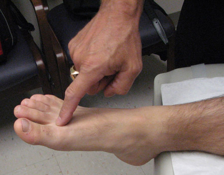
Evaluation of knee injury
First dorsal web space sensation testing (common peroneal nerve)
From the personal library of Dr LaPrade; used with permission
See this image in context in the following section/s:
Evaluation of knee injury
Hip internal rotation
From the personal library of Dr LaPrade; used with permission
See this image in context in the following section/s:
Evaluation of knee injury
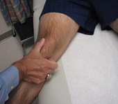
Palpation of prepatellar bursae
From the personal library of Dr LaPrade; used with permission
See this image in context in the following section/s:
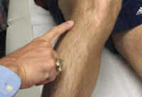
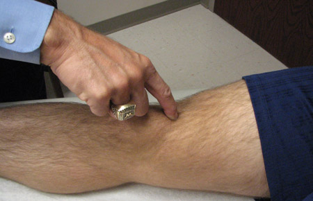
Evaluation of knee injury
Palpation of medial patellar fat pad
From the personal library of Dr LaPrade; used with permission
See this image in context in the following section/s:

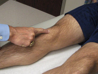
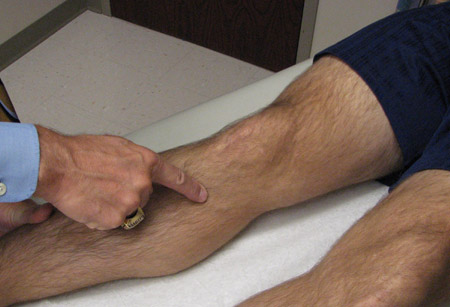
Evaluation of knee injury
Palpation of meniscotibial MCL
From the personal library of Dr LaPrade; used with permission
See this image in context in the following section/s:

Evaluation of knee injury
Dial test at 90° knee flexion in prone position
From the personal library of Dr LaPrade; used with permission
See this image in context in the following section/s:
Evaluation of knee injury
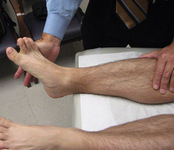
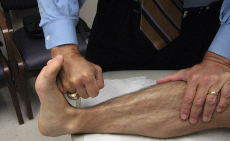
Dorsiflexion strength testing
From the personal library of Dr LaPrade; used with permission
See this image in context in the following section/s:
Evaluation of knee injury
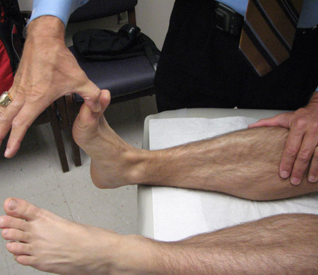
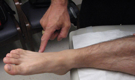
Extensor hallucis longus strength testing
From the personal library of Dr LaPrade; used with permission
See this image in context in the following section/s:
Evaluation of knee injury
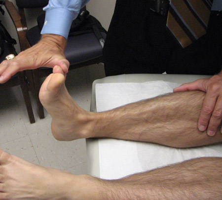
Small toe extensors strength testing
From the personal library of Dr LaPrade; used with permission
See this image in context in the following section/s:
Evaluation of knee injury
Log rolling test
From the personal library of Dr LaPrade; used with permission
See this image in context in the following section/s:
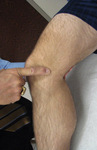
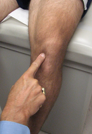
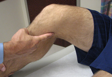
Evaluation of knee injury
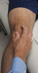
Palpation of superior pole of patella
From the personal library of Dr LaPrade; used with permission
See this image in context in the following section/s:

Evaluation of knee injury
Medial patellar translation
From the personal library of Dr LaPrade; used with permission
See this image in context in the following section/s:
Evaluation of knee injury
Palpation of pes anserine bursae
From the personal library of Dr LaPrade; used with permission
See this image in context in the following section/s:

Evaluation of knee injury
Rolling superior and inferior poles of patella within trochlear groove
From the personal library of Dr LaPrade; used with permission
See this image in context in the following section/s:
Evaluation of knee injury
Palpation of semimembranosus bursa
From the personal library of Dr LaPrade; used with permission
See this image in context in the following section/s:

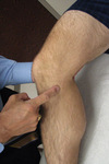
Evaluation of knee injury
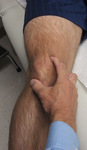
Palpation of inferior pole of patella
From the personal library of Dr LaPrade; used with permission
See this image in context in the following section/s:
Evaluation of knee injury
Quadriceps active test
From the personal library of Dr LaPrade; used with permission
See this image in context in the following section/s:
Evaluation of knee injury
Palpation of medial plica
From the personal library of Dr LaPrade; used with permission
See this image in context in the following section/s:
Evaluation of knee injury
Dial test at 90° knee flexion in supine position
From the personal library of Dr LaPrade; used with permission
See this image in context in the following section/s:
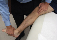
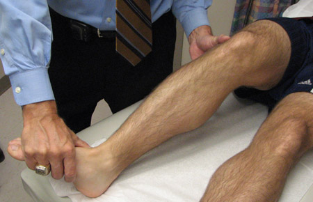
Evaluation of knee injury
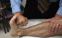
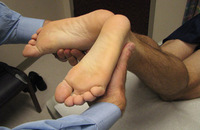
Foot inversion strength testing
From the personal library of Dr LaPrade; used with permission
See this image in context in the following section/s:

Evaluation of knee injury
Medial foot sensation (saphenous nerve)
From the personal library of Dr LaPrade; used with permission
See this image in context in the following section/s:

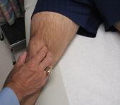
Evaluation of knee injury
Palpation of meniscofemoral MCL
From the personal library of Dr LaPrade; used with permission
See this image in context in the following section/s:

Evaluation of knee injury
Hip external rotation
From the personal library of Dr LaPrade; used with permission
See this image in context in the following section/s:

Evaluation of knee injury
Reverse pivot shift test
From the personal library of Dr LaPrade; used with permission
See this image in context in the following section/s:

Evaluation of knee injury
Dial test at 30° knee flexion in supine position
From the personal library of Dr LaPrade; used with permission
See this image in context in the following section/s:
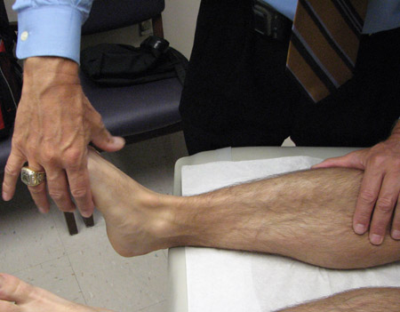
Evaluation of knee injury
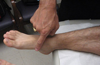
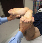
Foot eversion strength testing
From the personal library of Dr LaPrade; used with permission
See this image in context in the following section/s:

Evaluation of knee injury
Lateral patellar translation
From the personal library of Dr LaPrade; used with permission
See this image in context in the following section/s:
Evaluation of knee injury
Palpation of lateral patellar fat pad
From the personal library of Dr LaPrade; used with permission
See this image in context in the following section/s:
Evaluation of knee injury
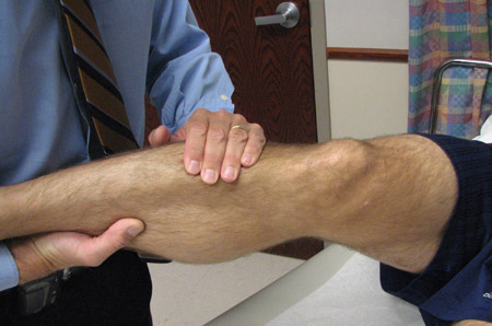
Hamstring strength testing
From the personal library of Dr LaPrade; used with permission
See this image in context in the following section/s:

Evaluation of knee injury
Rolling superior and inferior poles of patella within trochlear groove
From the personal library of Dr LaPrade; used with permission
See this image in context in the following section/s:
Evaluation of knee injury
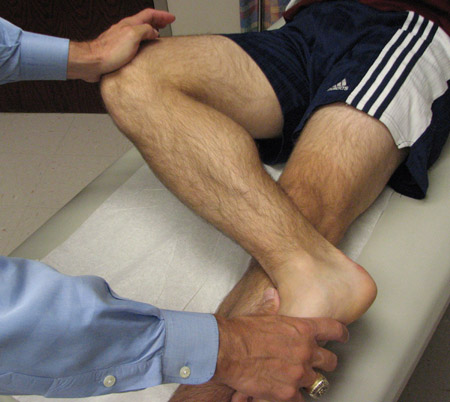
Lachman test
From the personal library of Dr LaPrade; used with permission
See this image in context in the following section/s:
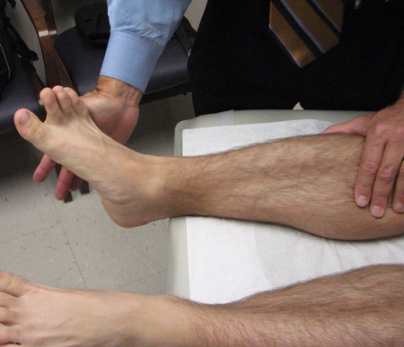
Evaluation of knee injury
Lateral foot sensation testing (superficial peroneal nerve)
From the personal library of Dr LaPrade; used with permission
See this image in context in the following section/s:

Evaluation of knee injury
Palpation of the biceps femoris bursae
From the collection of Dr LaPrade; used with permission
See this image in context in the following section/s:

Evaluation of knee injury
Quadriceps strength testing
From the personal library of Dr LaPrade; used with permission
See this image in context in the following section/s:
Evaluation of knee injury
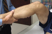
External rotation recurvatum test
From the personal library of Dr LaPrade; used with permission
See this image in context in the following section/s:

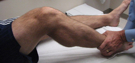
Evaluation of knee injury
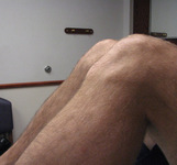
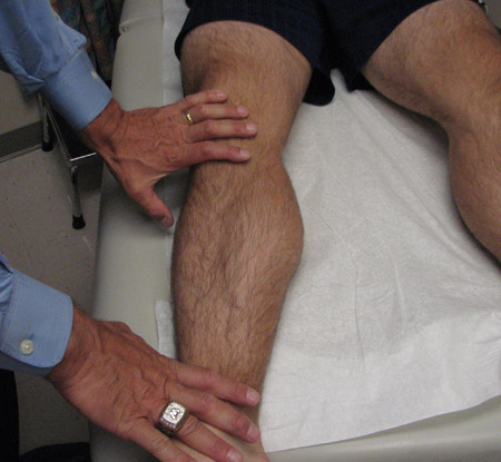
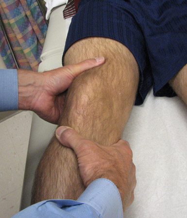
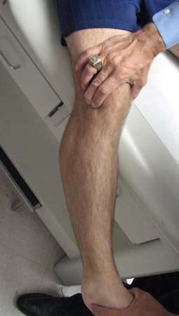
Deep flexion of the knee
From the collection of Dr LaPrade; used with permission
See this image in context in the following section/s:

Evaluation of knee injury
Posterior sag sign
From the personal library of Dr LaPrade; used with permission
See this image in context in the following section/s:

Evaluation of knee injury
Posterior drawer test
From the personal library of Dr LaPrade; used with permission
See this image in context in the following section/s:
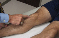
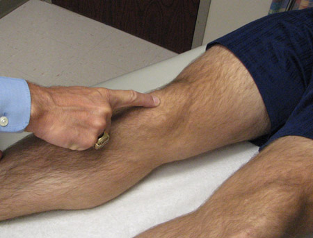
Evaluation of knee injury
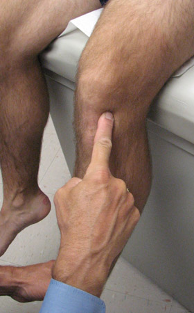
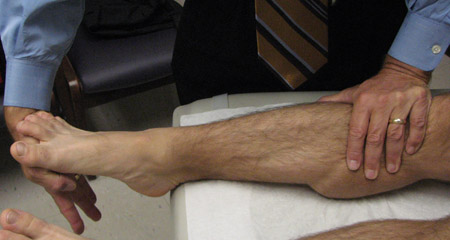
Palpation of tibial tubercle
From the personal library of Dr LaPrade; used with permission
See this image in context in the following section/s:
Evaluation of knee injury
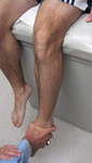
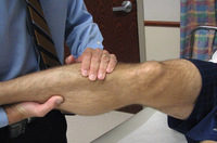
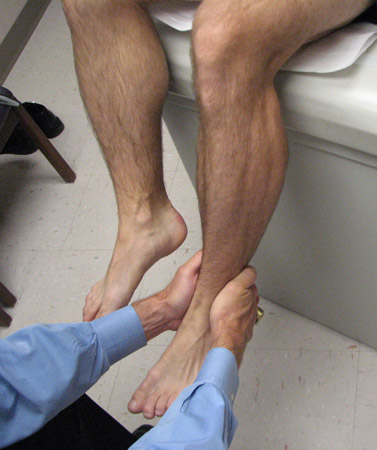
Plantar flexion strength testing
From the personal library of Dr LaPrade; used with permission
See this image in context in the following section/s:

Evaluation of knee injury
Dial test at 30° knee flexion in prone position
From the personal library of Dr LaPrade; used with permission
See this image in context in the following section/s:
Evaluation of knee injury
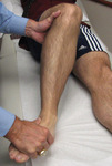
Varus stress test
From the personal library of Dr LaPrade; used with permission
See this image in context in the following section/s:
Use of this content is subject to our disclaimer