Images and videos
Images


Chronic pyelonephritis
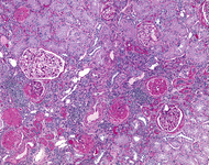
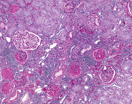
Chronic pyelonephritis: tubular damage, interstitial scarring, and fibrosis in chronic pyelonephritis
Courtesy of Jean L. Olson, MD, Department of Pathology, University of California, San Francisco, US
See this image in context in the following section/s:
Chronic pyelonephritis
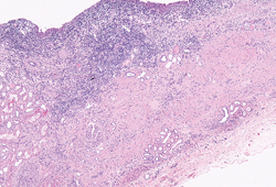
Medium-power microscopic view of dense polymorphonuclear infiltrates, showing chronic tubular atrophy, glomerulosclerosis, and surrounding areas of normal tubules and glomeruli
Courtesy of Jean L. Olson, MD, Department of Pathology, University of California, San Francisco, US
See this image in context in the following section/s:

Chronic pyelonephritis
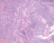
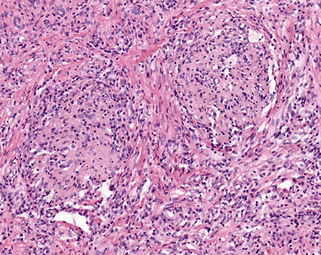
Xanthogranulomatous pyelonephritis: very low-power microscopic view showing extensive cellular infiltrate and granulomas. Note the marked destruction of the normal renal architecture
Courtesy of Jean L. Olson, MD, Department of Pathology, University of California, San Francisco, US
See this image in context in the following section/s:
Chronic pyelonephritis
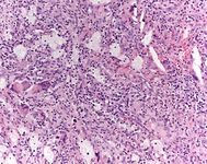
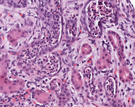
H&E stain: high-power view of granulomas found in xanthogranulomatous pyelonephritis
Courtesy of Jean L. Olson, MD, Department of Pathology, University of California, San Francisco, US
See this image in context in the following section/s:

Chronic pyelonephritis
Very high-power view of xanthoma cells, which are lipid-filled macrophages
Courtesy of Jean L. Olson, MD, Department of Pathology, University of California, San Francisco, US
See this image in context in the following section/s:
Chronic pyelonephritis
High-power view of polymorphonuclear cells in the renal tubules, showing damage to the tubules and red blood cell infiltration in the interstitial spaces
Courtesy of Jean L. Olson, MD, Department of Pathology, University of California, San Francisco, US
See this image in context in the following section/s:
Use of this content is subject to our disclaimer