Approach
Most patients with PDB are asymptomatic.[3] Patients who have symptoms are often initially misdiagnosed with another musculoskeletal disorder. Diagnosis is commonly incidental on imaging when the patient is being worked up for other diagnoses (e.g., to investigate renal disease), or on laboratory finding of isolated elevated alkaline phosphatase.[3] The diagnosis is confirmed by imaging studies.
Clinical findings
While most patients are asymptomatic, others may present with chronic pain in long bones and, less frequently, in facial areas.[3] The pain is typically continuous through the day and at rest, not related to positioning or loading, and is reported as aching or burning. Less frequently, pain may be secondary to pathologic fractures that occur as a result of weakened bones, most commonly the femur, hip, or tibia. In more severe cases, gross deformities of the affected bones can develop, including bowing of long bones or enlargement and thickening of skull and facial bones, which may present as visible frontal bossing or prognathism. Loosening of the teeth or malocclusion may occur when the facial bones are affected.[26] Increased local temperature may occur as a consequence of increased blood flow and metabolic activity.
Hearing loss is frequent in patients with PDB, and it is caused by cochlear damage more than otic nerve entrapment.[27]
In subjects with vertebral PDB, spinal stenosis is a common complication caused by expansion of the vertebral bodies and narrowing of the spinal canal, or by a blood-supply-steal mechanism.[3] Occasionally, patients may present with a pathologic vertebral compression fracture when the disease is localized to one or more vertebrae. Such patients may present with paresthesias and muscle weakness.[23][24][Figure caption and citation for the preceding image starts]: Clinical bowing of the tibia in Paget's disease (saber tibia)From the collection of Camilo Restrepo, Rothman Institute, Philadelphia, PA [Citation ends].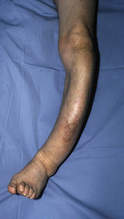
Initial imaging
Plain x-ray
Radiographic examination is central to the evaluation of PDB. X-rays are performed in all patients at the initial stage of evaluation.[28] The radiographs of pagetoid bone have a classical appearance.
The radiographic stages of the disease parallel the histologic stages. When osteoclast activity is present during the first stage of the disease, lytic changes can be observed on plain radiographs. This is most commonly seen in the skull and has been described as osteoporosis circumscripta cranii. In the long bones, this phase can be seen as an advancing V-shaped lytic lesion, which has been described as a flame edge or as having a blade-of-grass appearance.[29]
The second stage has no particular radiographic findings, but occasional fractures may be present, secondary to the disruption of the normal architecture by the pagetic process, which in turn compromises biomechanical competence of the affected bone. Over time, as the pagetic bone is stressed under weight-bearing and muscle-tension forces, it slowly deforms.
In the later stage, a sclerotic picture predominates. All three stages may be present in the same patient and the same bone at the same time. The characteristic thickened, coarse trabeculae seen on radiographs correspond histologically to a mosaic pattern of poorly organized lamellar bone.[23]
Bone scans (e.g., scintigram)
Technetium (Tc-bisphosphonate) scans are most commonly used as a screening tool to identify areas affected by the process, as not all pagetic sites may elicit local symptoms.[3][30] However, scintigraphic scans are not diagnostic.
Areas affected by PDB show as sites of intense uptake because of the high metabolic activity. However, in the first osteolytic stage, bone scans may be negative because of the low uptake of isotope.[29]
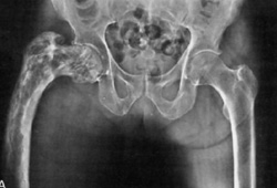
Serial Tc-bisphosphonate scans are not recommended even though treatment reduces radionuclide uptake in pagetic lesions.[28][31][Figure caption and citation for the preceding image starts]: X-ray of Paget's disease of proximal femurFrom the collection of Camilo Restrepo, Rothman Institute, Philadelphia, PA [Citation ends].
 [Figure caption and citation for the preceding image starts]: X-ray of Paget's disease of the tibiaFrom the collection of Camilo Restrepo, Rothman Institute, Philadelphia, PA [Citation ends].
Laboratory tests
All of the following tests are performed when a patient is suspected of having PDB.
Total serum alkaline phosphatase, a marker of bone formation, is an indicator of osteoblastic activity. Serum alkaline phosphatase is usually elevated in PDB, although patients with limited disease extension may have normal alkaline phosphatase (15%).[32] However, one study found normal levels of alkaline phosphatase in up to 86% of the patients.[33] When elevated, serum alkaline phosphatase can be followed to monitor response to treatment.[28] Other liver function tests should be measured to rule out liver disease as a possible cause of the increased alkaline phosphatase.
Bone-specific alkaline phosphatase is a more sensitive blood test for diagnosis, and can also be used as an index for treatment response, but availability may be limited.[34]
Serum procollagen 1 N-terminal peptide (P1NP, the amino-terminal propeptide of type 1 collagen) is a specific measure of bone formation that correlates well with activity on bone scintigraphy, but availability may be limited and/or cost prohibitive.[28] P1NP may be particularly useful in patients with monostotic or limited disease who may have normal serum alkaline phosphatase. P1NP can also be used as an index for treatment response.
Serum C-terminal propeptide of type 1 collagen (CTX), a measure of bone resorption activity, may be elevated in untreated PDB. Serum CTX can be used to monitor response to treatment as it decreases rapidly in response to bisphosphonate therapy.[28]
Serum 25-hydroxyvitamin D is measured to exclude vitamin D deficiency and osteomalacia as an alternative cause of elevated alkaline phosphatase.
Serum calcium measurements are recommended. Hypercalcemia, though rare, has been reported in patients with PDB, particularly after prolonged bed rest.
Further imaging
Computed tomography (CT) scans and magnetic resonance imaging (MRI) are used mostly for the evaluation of atypical or ambiguous presentations.[23][29] When patients present with neurologic involvement, imaging helps determine whether the neurologic symptoms are related to metabolic and physical bone changes in pagetoid bone.[35] Imaging is also used to investigate for possible malignant transformation. The pagetoid bone appears capacious and disorganized on cross-sectional studies such as CT and MRI scans.
Bone biopsy
The most sensitive and specific test for diagnosis is an undecalcified bone biopsy after tetracycline labeling, but this is rarely needed. In weight-bearing long bones such as the femur, diagnostic biopsy should be avoided because of the risk of fracture in an already compromised bone.[36] A bone biopsy is indicated if the diagnosis is still in doubt after the imaging and laboratory studies have been performed.
Use of this content is subject to our disclaimer