Images and videos
Images

Evaluation of traumatic brain injury, acute
Adult and pediatric GCS
From Dr Micelle J. Haydel; used with permission
See this image in context in the following section/s:
Evaluation of traumatic brain injury, acute
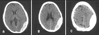
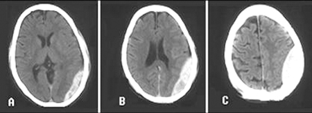
Epidural hematoma: CT brain scan showing lenticular-shaped hyperdensity between the dura mater and skull. (A–C) Same patient on different levels of the skull, (A) being the most caudal and (C) the most cranial
van Dijk GW. Practical Neurology. 2011 Feb;11(1):50-5
See this image in context in the following section/s:
Evaluation of traumatic brain injury, acute
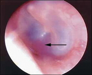
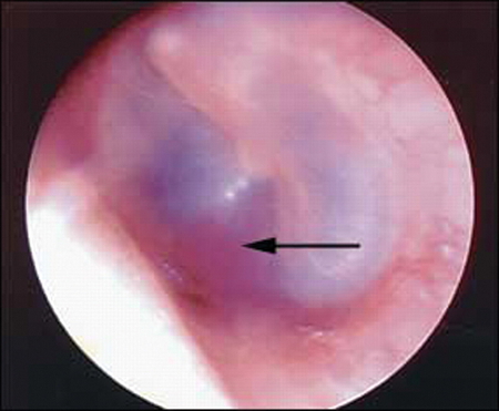
Hemotympanum: blood in the tympanic cavity of the middle ear (arrow)
van Dijk GW. Practical Neurology. 2011 Feb;11(1):50-5
See this image in context in the following section/s:

Evaluation of traumatic brain injury, acute
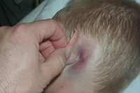
Battle sign: superficial ecchymosis over the mastoid process
van Dijk GW. Practical Neurology. 2011 Feb;11(1):50-5
See this image in context in the following section/s:
Videos
 Venepuncture and phlebotomy: animated demonstration
How to take a venous blood sample from the antecubital fossa using a vacuum needle.
Use of this content is subject to our disclaimer