Images and videos
Images


Evaluation of vaginal discharge
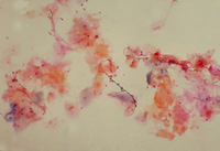
Vaginal smear identifying Candida albicans using a wet mount technique
CDC Image Library; Dr Stuart Brown
See this image in context in the following section/s:

Evaluation of vaginal discharge
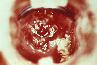
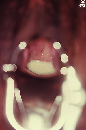
Cervicitis and vaginal discharge due to gonorrhea
CDC Image Library
See this image in context in the following section/s:

Evaluation of vaginal discharge
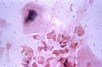
Vaginal smear identifying Candida albicans using Gram stain technique
CDC Image Library; Dr Stuart Brown
See this image in context in the following section/s:

Evaluation of vaginal discharge
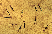
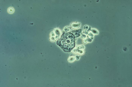
Micrograph of polymorphonuclear leukocytes and diplococci on a cervical smear
CDC Image Library; Joe Miller
See this image in context in the following section/s:
Evaluation of vaginal discharge
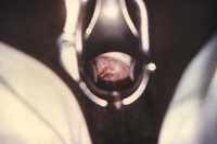
Trichomonasvaginitis with copious purulent discharge emanating from the cervical os
CDC Image Library
See this image in context in the following section/s:

Evaluation of vaginal discharge
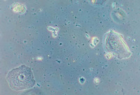
McCoy cell monolayer micrograph revealing intracellular Chlamydia trachomatis inclusion bodies; magnified 50x
CDC; Dr E. Arum, Dr N. Jacobs
See this image in context in the following section/s:
Evaluation of vaginal discharge
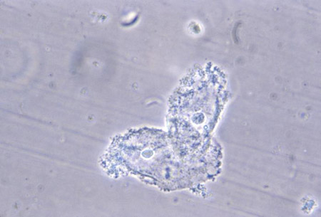
Phase contrast wet mount micrograph of a vaginal discharge revealing the presence of Trichomonas vaginalis protozoa
CDC Image Library
See this image in context in the following section/s:

Evaluation of vaginal discharge
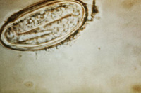
Enterobius vermicularis egg (human pinworm)
CDC Image Library
See this image in context in the following section/s:

Evaluation of vaginal discharge
Cervicitis due to herpes simplex virus; erosive inflammation with accompanying paracervical purulency is seen
CDC; Dr Paul Wiesner
See this image in context in the following section/s:
Evaluation of vaginal discharge
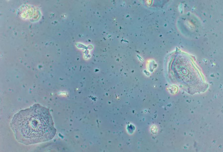
Photomicrograph revealing bacteria adhering to vaginal epithelial cells, known as clue cells
CDC Image Library; M. Rein
See this image in context in the following section/s:
Use of this content is subject to our disclaimer