Approach
Owing to the nature of this condition (i.e., an erect penis in the absence of sexual excitation), priapism is readily recognized clinically. Diagnosis should focus on the nature of the underlying type (i.e., ischemic, nonischemic, or recurrent) and the identification of contributory/predisposing conditions.
Clinical evaluation
When any patient presents with a prolonged erection, a detailed medical history and physical exam should be performed.[1] This should include:
Information about the duration of priapism
Existence of preceding factors (e.g., use of vasoactive drugs)
Any relieving maneuvers or clinical treatments used
Previous priapism episodes
Presence of pain
Existence of etiologic conditions or comorbidities (e.g., hemoglobinopathies), and
Erectile function status before the priapism episode.
Direct examination of the penis allows assessment of the extent of erection, degree of corporal body involvement (e.g., tricorporal priapism), and presence and severity of tenderness. In ischemic priapism, the penis and corpora cavernosa are rigid and tender to palpation. By contrast, in nonischemic priapism, the penis is not painful and is usually not rigid. Abdominal, perineal, and rectal exams may reveal signs of trauma, malignancy, or pelvic infection.[1] When spinal cord injury or lesion is suspected, a full neurologic exam may be indicated.[34]
Some patients may present with recurrent (sometimes called stuttering) priapism. Patients with stuttering priapism typically complain of painful episodes of priapism lasting <4 hours, which may occur weekly or every 2 weeks. Erections typically detumesce on their own, although in some cases penile edema and partial erection may remain.
Laboratory investigations
Complete blood count, white blood cell (WBC) differential, and platelet count should be ordered as initial tests in all patients with priapism to assess for any hematologic abnormalities or presence of an acute infection.[1][4] The WBC count may be raised, suggesting infection (e.g., pelvic abscess) or blood dyscrasia; anemia and increased reticulocyte count may be present in patients with sickle cell disease.
Sickle cell status should be ascertained in patients in whom this is uncertain or suspected. In these patients, rapid diagnosis can be obtained using a sickle-solubility test. This test detects any sickle hemoglobin (Hb) and therefore is positive in patients with both sickle cell trait and sickle cell disease. Subsequent confirmation of status requires Hb electrophoresis. Urine toxicology screening and coagulation profile may also be obtained.[1][4]
Penile diagnostics
After careful history taking, a cavernous blood gas analysis is useful in differentiating between ischemic and nonischemic priapism.[1] Blood aspirated from the corpus cavernosum offers an opportunity for direct visualization and evaluation of penile blood.[35] In patients with ischemic priapism, the aspirated blood is hypoxic and dark, whereas in nonischemic priapism, the blood is oxygenated and therefore bright red.
Cavernous blood gas results provide an immediate distinction between the classic divisions of priapism. Cavernous blood gas values for ischemic priapism typically show a pO₂ of <30 mmHg, pCO₂ of >60 mmHg, and pH of <7.25. Conversely, cavernous blood gas values for nonischemic priapism are pO₂ >90 mmHg, pCO₂ <40 mmHg, and pH 7.40, consistent with normal arterial blood at room air.[1][4]
For comparison, normal flaccid penis cavernous blood gas levels are similar to those of normal mixed venous blood at room air conditions (pO₂ 40 mmHg, pCO₂ 50 mmHg, and pH 7.35). [Figure caption and citation for the preceding image starts]: The technique of penile blood aspiration (corpora cavernosal needle placement for maximal corporal body irrigation)Arthur L. Burnett, MD, FACS [Citation ends].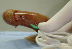
Imaging studies
Color duplex ultrasonography is not a first-line diagnostic test for priapism, but may be useful to differentiate between ischemic and nonischemic disease in a nonacute setting.[1] In ischemic priapism, minimal or absent blood flow is seen in the cavernosal arteries, as well as within the corpora cavernosa; patients with nonischemic priapism have normal to high blood flow velocities in the cavernosal arteries with evidence of blood flow in the corpora cavernosa.[1][2][32]
Ultrasonography may also show anatomic abnormalities, such as a cavernous arterial fistula or pseudoaneurysm, which would help confirm the diagnosis of nonischemic priapism. The study should be performed with the patient placed in lithotomy or frog leg position, to scan the perineum first and then the entire penile shaft.[2]
MRI may be considered to evaluate for corporal smooth muscle necrosis in patients with prolonged priapism being considered for penile prosthesis placement.[4] It is unlikely to have a role in the initial diagnosis of priapism.[1]
Penile arteriography has largely been supplanted by color duplex ultrasonography. However, it does have a role as part of an embolization procedure for nonischemic priapism.[4]
Use of this content is subject to our disclaimer