Images and videos
Images


Prion disease
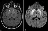
Changes in the basal ganglia seen in Creutzfeldt-Jakob disease. (A) Fluid-attenuated inversion recovery MRI and (B) diffusion-weighted MRI of the same patient demonstrate bilateral basal ganglia hyperintensities (arrows). There is also mild bilateral medial thalamus and pulvinar hyperintensity
From the personal collection of Dr M. Geschwind
See this image in context in the following section/s:
Prion disease
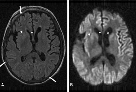
Diffuse cortical ribboning (arrows) seen on (A) diffusion-weighted imaging (DWI) and less so on (B) fluid-attenuated inversion recovery (FLAIR) MRI. Both sequences show cerebral cortex gyral hyperintensities
From the personal collection of Dr M. Geschwind
See this image in context in the following section/s:

Prion disease
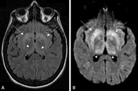
Bilateral medial thalamus and pulvinar hyperintensity (arrowheads) on (A) fluid-attenuated inversion recovery and (B) diffusion-weighted MRI in a patient with Creutzfeldt-Jakob. This patient also has significant basal ganglia hyperintensity on both sequences (arrows)
From the personal collection of Dr M. Geschwind
See this image in context in the following section/s:
Use of this content is subject to our disclaimer