Approach
Diagnosis is typically clinical. Impetigo usually presents a classic facial yellowish to golden crusting in a well individual.
History
Lesions are often asymptomatic, but occasionally are pruritic. They are usually not painful. There is often a history of contact with impetigo-infected peers at school or in playgroups. Lesions typically spread progressively from one site to another.
Exam
Vesicles are transient and are seldom seen; patients often present after this stage, with erosions that have a yellowish to golden crust on an erythematous base, with patchy distribution, often in the perioral and perinasal area, although they can occur anywhere on the body.
The streptococcal form tends to have thicker and darker crusts.
In resolving impetigo, the crusts usually dry and separate, leaving an erythematous base.
In nonhealing cases, there is elevation of the crust by the underlying mucopurulent exudate of active disease.
Involvement of mucous membranes is rare in the nonbullous form.
In the bullous form, bullae are often 2 cm in diameter or larger. They are initially clear and later become turbid. Buccal mucosa may be involved.
Severe disease, with systemic symptoms and signs such as lymphadenopathy and fever, may occur when large areas are affected.
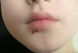
Suspicion of MRSA is raised in cases of spontaneous abscess or cellulitis or when lesions do not resolve with recommended initial antibiotic treatment.[Figure caption and citation for the preceding image starts]: Facial impetigo, yellow crust no longer visibleFrom the collection of Michael Freeman; used with permission [Citation ends].
 [Figure caption and citation for the preceding image starts]: Neonate with bullous impetigoFrom the collection of Michael Freeman; used with permission [Citation ends].
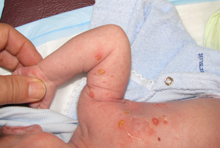 [Figure caption and citation for the preceding image starts]: Florid bullous impetigoFrom the collection of Michael Freeman; used with permission [Citation ends].
Laboratory
Usually, confirmatory tests are not necessary unless MRSA is suspected; if this is the case, confirm with appropriate swabs for bacteriologic culture.
Skin culture may also be prudent when the diagnosis is in doubt, or if the patient fails to respond to appropriate empiric therapy for impetigo.[1][24]
Use of this content is subject to our disclaimer