Approach
Acute burn management
Initial evaluation of burns should follow a systematic approach emphasizing patient stability, including early identification and control of airway and breathing problems, appropriate fluid resuscitation, recognition and treatment of associated injuries, and prompt consultation with burn specialist services where appropriate.[24] Limited burn-specific initial first aid should be provided. This includes:[37]
Cooling thermal burns with running water (up to 20 minutes; although optimal duration of cooling remains unclear, and guideline recommendations vary).[25][37]
Rinsing chemical burns on the skin or in the eyes with running water.
Covering a burn with a clean wet cloth or plastic cling wrap to protect it during transit to medical care (burns on an extremity should be covered rather than wrapped to allow for possible swelling).[25]
Do not deroof or aspirate blisters, as this may increase the risk of infection.
Outpatient burn management
Most burns can be managed in the outpatient setting by nonspecialists, but inadequately coordinated outpatient burn care can make for a frustrating and painful experience for the patient.[38] The key is careful patient selection and a well-rehearsed care plan.
Patients with smaller burns who have adequate support at home can generally be managed in the outpatient setting if this is deemed appropriate. Wounds of the face, ears, hands, genitals, and feet have a functional and cosmetic importance out of proportion to their wound size. In such cases, early specialty evaluation is advisable, unless the injuries are very superficial. Most burns selected for outpatient management are superficial and heal within 2 weeks. If this is not the case, patients may benefit from specialty evaluation.
Inpatient and specialist burn management
Patients who cannot take fluid by mouth, need burn resuscitation, potentially have inhalation injury, or cannot be managed in the outpatient setting should be admitted for inpatient care. Where possible, consult with a specialist burns center and arrange transfer as appropriate.[39][40]
Some patients initially managed in the clinic setting will subsequently require admission. Reasons prompting admission include:
Increased pain and anxiety
Inability to keep scheduled appointments
Delayed healing
Signs of infection
Wound that appears deeper than initially estimated. Burn depth is commonly underestimated during the first days after injury
Serious burns are most effectively and least expensively managed in organized programs focused on burn care. An increasing body of data support the efficacy of concentration of serious burns in regional programs.[41]
The American Burn Association (ABA) has established criteria for determining which patients require referral to a specialized burn center, but local resources and practice patterns should be taken into account. The ABA states that the following burn injuries should be referred:[41]
Partial-thickness burns of >10% total body surface area
Burns that involve the face, hands, feet, genitalia, perineum, or major joints
Third-degree burns in any age group
Electrical burns, including lightning injury
Chemical burns
Inhalation injury
Burn injury in patients with pre-existing medical disorders that could complicate management, prolong recovery, or affect mortality
Burned children in hospitals without qualified personnel or equipment for the care of children
Burn injury in patients who will require special social, emotional, or rehabilitative intervention
Any patients with burns and concomitant trauma (such as fractures) in which the burn injury poses the greatest risk of morbidity or mortality. In such cases, if the trauma poses the greater immediate risk, the patient's condition may be stabilized initially in a trauma center before transfer to a burn center. Physician judgment will be necessary in such situations and should be in concert with the regional medical control plan and triage protocols.
An expert consensus panel has proposed updating and extending the original ABA referral criteria to include the following additional referral criteria/considerations:[42]
Full-thickness burns ≥ 5% TBSA burned.
Children and older adults (>55 years of age). These patients may benefit from referral to a burn center to access the multidisciplinary team resources, even when TBSA burned is less than 10% (partial or full thickness).
Smaller burns should be followed up in burn center outpatient settings as soon as possible after injury, and preferably within a week.
Consider telemedicine consultations as an alternative to immediate transfer or outpatient referral for selected patients.
Internationally, burn center transfer criteria vary and may depend on local resources and/or configuration of specialist burn services.[43] In UK practice, for example, specialized burn services are designated as burn facilities, burn units, and burn centers according to the level of injury complexity they manage. A burn facility offers inpatient care for noncomplex burn injuries at the level of a standard plastic surgical floor (criteria include burns of TBSA 2% to 4.9%). Burn units will accept patients requiring higher levels of care (including TBSA burned ≥5%), while burn centres accept the most severe and complex cases (e.g., TBSA burned ≥30% for adults and older children, or ≥15% if under 1 year old).[40]
A survival benefit has been demonstrated for patients with serious burns if they are managed in a dedicated high-volume burn center.[44]
A number of nonburn medical and surgical conditions (e.g., frostbite, Stevens-Johnson syndrome/TENS, and necrotizing soft-tissue infection) require the same specialized resources as burns. These conditions are increasingly recommended for referral to burn units for initial assessment and care.[42][45]
Outpatient treatment: wound care and inspection
Most minor burns can be managed in an outpatient setting. Initial treatment consists of wound cooling, cleaning, and dressing. Pain management and tetanus prophylaxis are important. Access for regular follow-up through the healing period is important. The frequency of follow-up will vary with a number of wound-related and other factors, including:
Wound severity
Patient age
Patient comfort
Family competence
Availability of community nursing resources
All patients should be told to return immediately if they have problems, questions, or signs of infection prior to their scheduled appointments.
Cleaning
Clean burn wounds with lukewarm tap water and a bland soap
Topical antibiotic prophylaxis
Systemic antibiotic prophylaxis is not routinely recommended as prior studies demonstrated no compelling benefit.[46] Topical prophylaxis remains a common practice, but it may not be necessary as supportive data is weak.
Topical agents range from aqueous solutions to antibiotic-containing ointments and debriding enzymes. Very little efficacy data exist.[47] Empiric practices are poorly supported. There are a great number of approaches to the care of small burns.[48] Common sense and regular reassessment of wounds as they heal are central to successful treatment.
Silver is an excellent antiseptic and there is a long history of its use in burn dressings despite a relative lack of evidence for efficacy.[49] [
 ]
It is applied in several forms, including silver sulfadiazine cream, aqueous silver nitrate solution, and dressings containing nanocrystalline silver.[50] Topical silver sulfadiazine is commonly used, as it is painless on application and has a broad spectrum of antibacterial activity, although some in vitro evidence suggests it may slow epithelialization to a modest degree and some guidelines advise against its use.[37][49][51][52]
]
It is applied in several forms, including silver sulfadiazine cream, aqueous silver nitrate solution, and dressings containing nanocrystalline silver.[50] Topical silver sulfadiazine is commonly used, as it is painless on application and has a broad spectrum of antibacterial activity, although some in vitro evidence suggests it may slow epithelialization to a modest degree and some guidelines advise against its use.[37][49][51][52]There is limited experience with debriding enzymes in the outpatient setting, with no evolved standard of care. In some centers they play an important role, but in others their utility is limited by expense.
Superficial burns can be treated with viscous antibacterial ointments containing low concentrations of various antibiotics.
Wounds around the eyes can be treated with topical ophthalmic antibiotic ointments.
Treat deeper ear burns with mafenide, as it is the only agent that will penetrate relatively avascular cartilage. This is important as infection of the cartilaginous skeleton of the external ear can cause significant deformity.
Dressings
Simple gauze wraps minimize soiling of clothing and protect the wound from trauma.
Wound membranes are increasingly popular and effective, providing pain control, prevention of wound desiccation, and reduction of wound colonization. They help to create a moist wound environment with a low bacterial density, and are generally intended for use on selected clean superficial wounds and donor sites.[52] They should be used with caution, as deeper wounds placed in membrane dressings may become septic if they are not carefully monitored. Many will release silver for several days, reducing bacterial proliferation.[53][54]
In the first days immediately after burning, when wound depth is unclear, topical agents are often ideal. When it is clear that the wound is clean and superficial, a transition to membranes may be appropriate. The experience and comfort level of the treating physician is a major consideration when contemplating the routine use of membrane dressings.
Dressing changes
The wound should be kept generally clean and regularly inspected for infection.
Outpatient burns tend to be small and superficial, presenting a relatively low risk of infection, so clean rather than sterile technique is reasonable.
Accumulated exudate and topical medications should be gently cleansed with lukewarm tap water and a bland soap.
Soaking adherent dressings prior to removal will minimize any pain associated with their removal.
Gently cleanse wounds with a gauze or clean washcloth, inspect for any sign of infection, pat dry with a clean towel, and redress.
Inspection for infectious complications is important: the patient and family should be advised to return promptly if they notice erythema, swelling, increased tenderness, lymphangitis, odor, or drainage.
Wound inspection and cleansing will cause many patients significant anxiety and pain; some will benefit from an oral opioid given 30 to 60 minutes before a dressing change.
The interval between dressing changes will vary, but most small burns are adequately managed with a daily cleansing and a dressing change.
If wounds are clean and superficial and if surgery is not needed, wound check frequency can be reduced, or a membrane dressing can be applied.
Tetanus immunization is recommended in the absence of a history of adequate immunization.
Scar management
Scar management is an essential aspect of comprehensive burn care.
Unfortunately, grading of scars is not uniformly done, although a variety of scales do exist.[55] Scar evaluation remains a largely subjective practice.
In long-term scar management, pressure garments have been promoted for many years, although a meta-analysis failed to demonstrate reliable benefit. It may be that further study of subgroups with better scar measurement tools may demonstrate a benefit, but, for now, use is likely to remain program-dependent and anecdote-based.[56]
Early experiences with tuneable dye and fractional carbon dioxide lasers have been encouraging; however, controlled data do not exist.[57] Laser interventions seem to work well only when used as part of a multimodality scar management program.[58]
Inpatient treatment: fluid resuscitation, wound care, critical care, and surgery
Care of severe burns can be divided into 4 phases:[59]
Initial evaluation and resuscitation
Initial excision and biologic closure
Definitive wound closure
Rehabilitation and reconstruction
The phases overlap, and intensive care plays an important role in the first 3 phases.
1. Initial evaluation and resuscitation
Usually the first 24-72 hours after injury.
Document the full extent of the injury (including nonburn trauma) and perform fluid resuscitation.
Patients with airway involvement or major burns generally require intubation and mechanical ventilation, although intubation should be done selectively.[25][60][61][Figure caption and citation for the preceding image starts]: Airway edemaFrom Dr Sheridan's personal collection [Citation ends].

Carbon monoxide intoxication is best treated acutely by effective ventilation with 100% oxygen, but can be associated with delayed neurologic sequelae. Hyperbaric oxygen treatment is appropriate in selected stable patients with serious exposures, but it is not indicated for routine wound healing.[62]
If burns involve >20% of the body surface, reduced capillary integrity becomes clinically important, with a resulting need for fluid resuscitation.[24][25][63] Capillary integrity is typically restored at approximately 24 hours. Any of the several burn formulas available may be used to initiate resuscitation; however, none can be assumed to be accurate in an individual patient. Bedside titration of infusions, based on physiologic endpoints, is important. Gentle titration is advised: for example, if the urine output falls to 0.25 mL/kg/hour, consider increasing the infusion rate by 10% and reassessing in 60 minutes, rather than infusing a large bolus of fluid. The Parkland formula is often used, and suggests 4 mL/kg/% burn over the first 24 hours, half in the first 8 hours. Young children should receive 5% dextrose in lactated Ringer solution at a maintenance rate to ensure they do not develop hypoglycemia.
While all burn resuscitation formulae are meant to guide only the initial rate of fluid infusion, the choice of formula may be important. Some studies suggest that use of the Modified Brooke formula or alternative approaches may adequately resuscitate patients while limiting resuscitation volumes, compared to the widely-used Parkland formula.[64] The Modified Brooke formula suggests 2 mL/kg/% burn (% TBSA), where half of the total fluid requirement is given in the first 8 hours, and the other half is given in the next 16 hours.[24] The rate of fluid administration is adjusted based on the patient's urine output. During the second 24 hours, 0.33 to 0.5 mL/kg/% TBSA burned of colloid plus D5W is given to maintain urine output.[65] The American Burn Association (ABA) Clinical Practice Guideline (CPG) recommends 2 mL/kg/% TBSA burn for initial fluid resuscitation in adults with burns ≥20% TBSA in order to reduce resuscitation fluid volumes.[64]
Lactated Ringer solution is generally administered to burn patients, as specified in the Parkland formula.[63] However, the evidence for choice of crystalloid fluids for critically ill patients in general is conflicting, with very little good data specific to burn resuscitation.[63][66][67][68]
The role of colloid in burn resuscitation remains controversial. Many clinicians advise inclusion of colloid (generally albumin) in burn resuscitation when burns are large to reduce anasarca (severe, generalized interstitial fluid accumulation) despite evidence to the contrary from a meta-analysis and conflicting recommendations from some guidelines.[69][70][71] The ABA CPG advises to consider giving albumin in burn patients with >20% TBSA in the first 24 hours of burn resuscitation to improve urinary output and to reduce the total volume of resuscitation fluids.[64] The strength of this recommendation is greater for patients with larger burns and weaker for patients with smaller burns. As studies are also conflicting as to whether to start albumin before or after the first 12 hours, the ABA suggests in situations where albumin is part of the general resuscitation plan, versus in an acute rescue situation, that it is preferable to start the albumin after the first 12 hours until more is known.[64] Solutions containing hydroxyethyl starch (HES) are not recommended due to their increased risk of adverse outcomes including kidney injury and death, particularly in critically ill patients, and their use has been suspended in Europe.[24][63][72][73]
Even patients with massive burns can have a good outcome if managed in a comprehensive burn program. Fluid resuscitation becomes increasingly challenging as burn size increases. War-time experience has added to our understanding of the resuscitation needs of the very severely injured.[74]
Even during resuscitation, it is important to ensure attention is paid to patient comfort. Pain and anxiety can have adverse physiologic and emotional consequences.[75] Successful early pain control can enhance important aspects of long-term outcome.[76]
Heparin has been proposed as an adjunct to care of burns, but it is not part of the standard of care and data are not convincing.[77][78]
Tetanus immunization should be updated in patients with wounds deeper than a superficial partial-thickness burn.
2. Initial excision and biologic closure
Identification and removal of large areas of full-thickness burn before wound sepsis and systemic inflammation develop. This should be done using staged hemostatic and minimally ablative techniques.[79]
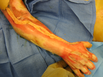
Near- or completely circumferential burns should be identified for special monitoring and possible escharotomy:[24][25][Figure caption and citation for the preceding image starts]: Circumferential burnFrom Dr Sheridan's personal collection [Citation ends].
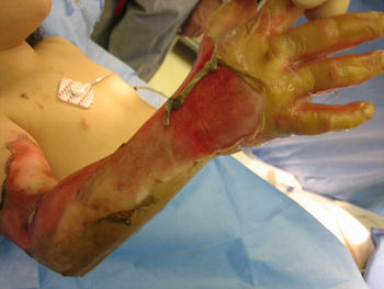
Burns of this sort on the torso can interfere with ventilation or even contribute to intra-abdominal hypertension.
On extremities, such burns may cause limb-threatening ischemia within 12-24 hours.
Escharotomy can decompress such problems and can be done with coagulating electrocautery; anesthesia or sedation is generally required in children.
When performing escharotomy it is important not to damage uninjured skin or superficial neurovascular structures.
[Figure caption and citation for the preceding image starts]: EscharotomyFrom Dr Sheridan's personal collection [Citation ends].
Ideally, close wounds with autograft. Temporary wound membranes can be useful for large wounds. This strategy changes the natural history of the injury from inevitable systemic sepsis and inflammation to a more controlled wound-closure situation.
Amniotic membrane can be an accessible and effective temporary membrane, but blood-borne infectious disease screening remains a concern and should be considered.[80]
The role of antibiotic prophylaxis during acute burn surgery remains unclear.
3. Definitive wound closure
Duration varies depending on wound size and complexity.
The objective is to replace any temporary membranes with autograft and to close small complex wounds, such as on the hands and face.
May take many weeks if donor sites are severely limited.
Intensive care is an important component of the first 3 phases of care. Ideally, an embedded intensive care unit is part of the burn program, so that coordination between the medical and surgical needs of the patients is seamless. A burn critical care capability can be organized in various ways, but must always foster a strong collaboration between the surgical, medical, nursing, and other disciplines.[81]
Deep venous thrombosis is a risk in all injured patients. There are few studies of this in burn patients to support a specific approach. Each unit should develop its own policy for monitoring, prophylaxis, and treatment.[82]
4. Rehabilitation and reconstruction
This is the longest phase of care.
Ideally, begins with early ranging and splinting, and antideformity positioning.
As wounds are closed and patients moved from intensive care, passive and active motion and strengthening become important.
Scar management and emotional support are extremely useful for most patients.
Burn reconstructive procedures are ideally planned as soon as functional or aesthetic deformities hinder further recovery.
Long-term follow-up is essential to optimize recovery, particularly for those with larger injuries.[83] This includes support of the family group.[84] Patient and family education efforts enhance understanding and participation in aftercare needs.[85]
Pruritus can be a persistent discomfort in the first months after wound closure and should be addressed with nonpharmacologic as well as pharmacologic means.[86]
Planning for long-term plastic and reconstructive surgery needs should be considered in disaster scenarios, as this need will continue after the flurry of initial activity and attention.[87]
Attention to pain and anxiety are essential in all phases of care. This is usually done by infusion of opioids and benzodiazepines (e.g., morphine sulfate and midazolam). Each unit should establish their own protocols and dosing regimens. Nonpharmacologic therapies, such as music therapy, can be useful in selected patients. Virtual reality is an innovative, new, nonpharmacologic, noninvasive analgesic technique. Although only few studies are available, positive initial experiences have been reported and a systematic review found it to be an effective adjunct for treatment of pain during wound dressing changes and physical therapy.[88][89]
Burn patients typically remain alert and oriented even with major burns, and can remember events at the time of the injury and for several hours afterward. Health care providers should be sensitive to the variable emotions experienced by burn patients and their families at all stages of treatment, and consider the psychosocial needs of the survivor during and following hospitalization and rehabilitation.[24]
Treatment of wound infection
Regular monitoring of burn wounds allows for the early recognition of infection. All suspected burn infections require aggressive management, which may include admission, intravenous antibiotics, observation, and surgery excision if wounds are deep.
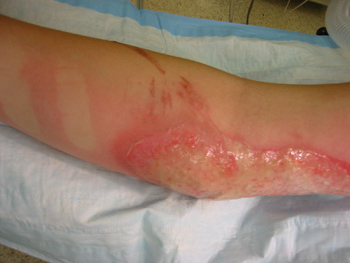
Burn wound cellulitis responds readily to antibiotics in most cases. [Figure caption and citation for the preceding image starts]: CellulitisFrom Dr Sheridan's personal collection [Citation ends].
Burn impetigo is usually associated with Staphylococcus aureus and Streptococcus pyogenes and is particularly common in burns of the scalp. Treatment requires wound cleansing, which often mandates shaving of nearby hair-bearing areas, and grafting of full-thickness areas.
Treat open burn-related surgical wound infections with debridement of necrotic and infected material with delayed wound closure.
Invasive burn wound infection is a serious problem, usually addressed by excision, and systemic and topical antibiotics.
Use of this content is subject to our disclaimer