Etiology
Burn injuries are caused by a number of mechanisms.[6]
Thermal burns:
Caused by heat, from hot liquids, flame, or contact with heated objects
In young children, about 70% of burns caused by scalding from hot liquids
In older children and young working adults, flame injuries are more likely
In older adults, scalds and cooking accidents are most common.
Electrical burns:
Caused by low-, intermediate-, and high-voltage exposures, causing a variety of local and systemic injuries.
Chemical burns:
Caused by exposure to industrial or household chemical products.
Nonaccidental burns:
Approximately 20% of burns in younger children involve abuse or neglect.
Pathophysiology
Burn injuries can result in local and systemic responses.
Local response
Involves the coagulation of injured tissue, and to some degree incites progressive microvascular reactions in the surrounding dermis.[7]
In animal models, the secondary injury caused by these microvascular changes has been truncated by a variety of administered substances, but none has been demonstrated to be clinically useful.[8]
Systemic response
As burns become larger than about 20% of the total body surface area (TBSA), a systemic response ensues, driving fluid loss and release of vasoactive mediators from the injured tissue. Clinically this results in early capillary leak, interstitial edema, and organ dysfunction.[9][10]
In well-resuscitated patients, this physiology will self-extinguish and be replaced by a hypermetabolic response, with a near doubling of cardiac output and resting energy expenditure over the next 24 to 48 hours. The magnitude of this response, peaking in those with injuries of 60% or more TBSA, is as high as twice the normal basal metabolic rate.[11] Accelerated gluconeogenesis, insulin resistance, and increased protein catabolism are associated with this response and have major implications for subsequent support of burn patients. The mechanism is not well understood but it is assumed to involve a combination of factors including a change in hypothalamic function; increased glucagon, cortisol, and catecholamine secretion; deficient gastrointestinal barrier function with translocation of bacterial byproducts; bacterial contamination of the burn wound with systemic release of similar products; and some element of enhanced heat loss via transeschar evaporation of fluid. Nutritional support of this physiologic response is essential. Numerous efforts to modify this process pharmacologically have proven less routinely effective.[12] Growth hormone has been advocated in seriously burned children, but data are not compelling and this is not widely practiced at present.[13] [
 ]
Data suggest that use of anabolic steroids may favorably influence burn physiology while also being less expensive, but this practice has not been broadly adopted either.[14]
]
Data suggest that use of anabolic steroids may favorably influence burn physiology while also being less expensive, but this practice has not been broadly adopted either.[14]The subsequent natural history of burns is driven by the wound. A burn wound is initially clean but is rapidly colonized by endogenous bacteria. As these bacteria multiply, proteases liquefy the eschar, which then separates, leaving a bed of granulation tissue or healing burn depending on the depth of the injury. In healthy patients with small burns this septic process is often well tolerated. However, when injuries are larger, systemic infection results, resulting in poor survival of patients with burns involving >40% TBSA managed without early wound excision.[15]
Classification
Standard clinical classification of burns according to depth
First-degree burns:
Erythema involving the epidermis only
Usually dry and painful
Typical of severe sunburn.
Second-degree burns:
Superficial partial-thickness burns involving the epidermis and upper dermis
Deep partial-thickness burns involving the epidermis and dermis
Usually wet and painful
Typical of scalding injury.
Third-degree burns:
Full-thickness burns involving the epidermis and dermis and damage to appendages
Usually dry and insensate
Typical of flame or contact injury.
Fourth-degree burns:
Involve underlying subcutaneous tissue, tendon, or bone
Typical of high-voltage electrical injury.[Figure caption and citation for the preceding image starts]: First-degree burnFrom Dr Sheridan's personal collection [Citation ends].
 [Figure caption and citation for the preceding image starts]: Second-degree burnFrom Dr Sheridan's personal collection [Citation ends].
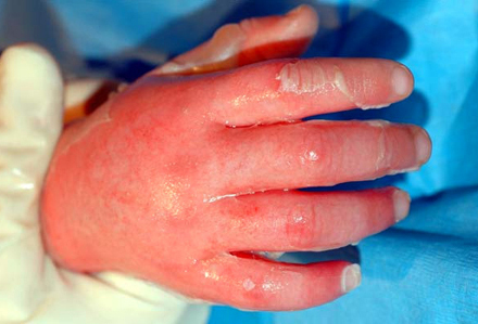 [Figure caption and citation for the preceding image starts]: Third-degree burnFrom Dr Sheridan's personal collection [Citation ends].
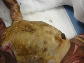 [Figure caption and citation for the preceding image starts]: Fourth-degree burnFrom Dr Sheridan's personal collection [Citation ends].
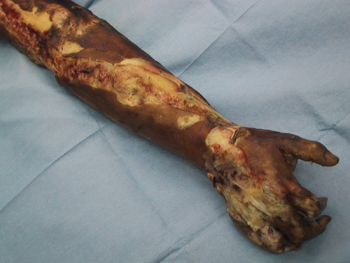
Use of this content is subject to our disclaimer