Images and videos
Images
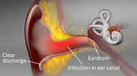
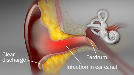
Otitis externa
Diagram of acute otitis externa
Created by the BMJ Knowledge Centre
See this image in context in the following section/s:
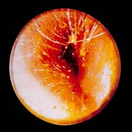
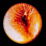
Otitis externa
Swollen ear canal, almost completely closed due to acute otitis externa
From the collection of Dr Richard Buckingham; used with permission
See this image in context in the following section/s:

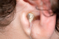
Otitis externa
White purulent debris can be seen at the external auditory meatus
Barry V et al. BMJ 2021;372:n714; used with permission
See this image in context in the following section/s:

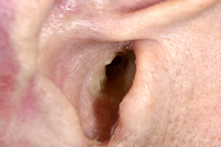
Otitis externa
The ear canal is narrowed, making it appear more slit-like, with white debris sitting on the canal wall
Barry V et al. BMJ 2021;372:n714; used with permission
See this image in context in the following section/s:
Videos
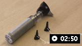 How to examine the ear
How to perform an examination of the ear.
Use of this content is subject to our disclaimer