Diagnosis is usually clinical, with patients presenting with rapid onset of symptoms.[3]Hirsch BE. Infections of the external ear. Am J Otolaryngol. 1992 May-Jun;13(3):145-55.
http://www.ncbi.nlm.nih.gov/pubmed/1626615?tool=bestpractice.com
History and physical exam
National guidelines state that a diagnosis of acute otitis externa (AOE) requires the presence of rapid onset (generally within 48 hours) of symptoms within the past 3 weeks, coupled with signs of ear canal inflammation.[1]Rosenfeld RM, Schwartz SR, Cannon CR, et al. American Academy of Otolaryngology-Head and Neck Surgery Foundation. Clinical practice guideline: acute otitis externa. Otolaryngol Head Neck Surg. 2014 Feb;150(1 suppl):S1-24. [Erratum in: Otolaryngol Head Neck Surg. 2014 Mar;150(3):504].
https://aao-hnsfjournals.onlinelibrary.wiley.com/doi/10.1177/0194599813517083
http://www.ncbi.nlm.nih.gov/pubmed/24491310?tool=bestpractice.com
Symptoms of ear canal inflammation include ear pain (which can be severe), itching, and fullness, with or without decreased hearing or pain in the ear canal and temporomandibular joint intensified by jaw motion. Signs of ear canal inflammation include tenderness over the tragus, pinna, or both.[1]Rosenfeld RM, Schwartz SR, Cannon CR, et al. American Academy of Otolaryngology-Head and Neck Surgery Foundation. Clinical practice guideline: acute otitis externa. Otolaryngol Head Neck Surg. 2014 Feb;150(1 suppl):S1-24. [Erratum in: Otolaryngol Head Neck Surg. 2014 Mar;150(3):504].
https://aao-hnsfjournals.onlinelibrary.wiley.com/doi/10.1177/0194599813517083
http://www.ncbi.nlm.nih.gov/pubmed/24491310?tool=bestpractice.com
Manipulation of the ear canal is usually painful. The skin of the external auditory canal has variable degrees of diffuse edema, erythema, and swelling. There may be otorrhea or cellulitis of the pinna and adjacent skin. Otoscopy is recommended to examine the state of the tympanic membrane.
Sometimes the canal is very swollen, and this obscures the examination of the tympanic membrane. Variable amounts of drainage and debris will be seen on otoscopic ear exam. The tympanic membrane may be erythematous. In certain instances, cervical lymphadenopathy may be present.
[Figure caption and citation for the preceding image starts]: Swollen ear canal, almost completely closed due to acute otitis externaFrom the collection of Dr Richard Buckingham; used with permission [Citation ends]. [Figure caption and citation for the preceding image starts]: White purulent debris can be seen at the external auditory meatusBarry V et al. BMJ 2021;372:n714; used with permission [Citation ends].
 [Figure caption and citation for the preceding image starts]: The ear canal is narrowed, making it appear more slit-like, with white debris sitting on the canal wallBarry V et al. BMJ 2021;372:n714; used with permission [Citation ends].
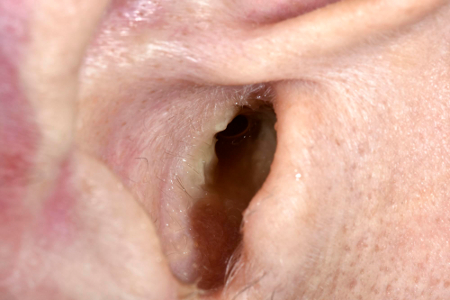
Pneumatic otoscopy and/or tympanometry
Pneumatic otoscopy and tympanometry can be performed to aid in the diagnosis.[1]Rosenfeld RM, Schwartz SR, Cannon CR, et al. American Academy of Otolaryngology-Head and Neck Surgery Foundation. Clinical practice guideline: acute otitis externa. Otolaryngol Head Neck Surg. 2014 Feb;150(1 suppl):S1-24. [Erratum in: Otolaryngol Head Neck Surg. 2014 Mar;150(3):504].
https://aao-hnsfjournals.onlinelibrary.wiley.com/doi/10.1177/0194599813517083
http://www.ncbi.nlm.nih.gov/pubmed/24491310?tool=bestpractice.com
Pneumatic otoscopy will demonstrate normal tympanic membrane movement, which may be absent in patients with associated acute otitis media. Similarly, in patients with AOE, tympanometry will be normal but will show a flat tracing (type B) in patients with associated acute otitis media. Tympanometry may cause discomfort and pain in patients with AOE.
Culture and microscopy
Ear cultures are obtained mainly from patients who fail to improve with medical therapy. Cultures are usually unnecessary on initial visit or at the time of diagnosis but can be obtained if desired.[3]Hirsch BE. Infections of the external ear. Am J Otolaryngol. 1992 May-Jun;13(3):145-55.
http://www.ncbi.nlm.nih.gov/pubmed/1626615?tool=bestpractice.com
The most commonly cultured organisms are Pseudomonas and Staphylococcus species.[1]Rosenfeld RM, Schwartz SR, Cannon CR, et al. American Academy of Otolaryngology-Head and Neck Surgery Foundation. Clinical practice guideline: acute otitis externa. Otolaryngol Head Neck Surg. 2014 Feb;150(1 suppl):S1-24. [Erratum in: Otolaryngol Head Neck Surg. 2014 Mar;150(3):504].
https://aao-hnsfjournals.onlinelibrary.wiley.com/doi/10.1177/0194599813517083
http://www.ncbi.nlm.nih.gov/pubmed/24491310?tool=bestpractice.com
Negative cultures are sometimes obtained in patients who are on antibiotic treatment, whether topical or systemic. Cultures positive for fungal species are found in patients with fungal otitis externa.
Microscopy of exudate/debris from the ear canal may reveal evidence of fungal infection. White filamentous hyphae are seen in fungal otitis externa (otomycosis). The presence of black spores indicates Aspergillus niger as the causative organism.[1]Rosenfeld RM, Schwartz SR, Cannon CR, et al. American Academy of Otolaryngology-Head and Neck Surgery Foundation. Clinical practice guideline: acute otitis externa. Otolaryngol Head Neck Surg. 2014 Feb;150(1 suppl):S1-24. [Erratum in: Otolaryngol Head Neck Surg. 2014 Mar;150(3):504].
https://aao-hnsfjournals.onlinelibrary.wiley.com/doi/10.1177/0194599813517083
http://www.ncbi.nlm.nih.gov/pubmed/24491310?tool=bestpractice.com
[3]Hirsch BE. Infections of the external ear. Am J Otolaryngol. 1992 May-Jun;13(3):145-55.
http://www.ncbi.nlm.nih.gov/pubmed/1626615?tool=bestpractice.com
Radiology
Computed tomography (CT) scans of the temporal bone with and without contrast are recommended in patients who have persistent severe ear pain and fullness despite adequate medical therapy with topical and oral antibiotics. This is to rule out necrotizing otitis externa. Clinical features that would suggest a need for a CT scan include pain that is disproportionate to the clinical findings and patients with granulation tissue along the floor of the external auditory canal, especially in patients with diabetes or those who are immunocompromised.[1]Rosenfeld RM, Schwartz SR, Cannon CR, et al. American Academy of Otolaryngology-Head and Neck Surgery Foundation. Clinical practice guideline: acute otitis externa. Otolaryngol Head Neck Surg. 2014 Feb;150(1 suppl):S1-24. [Erratum in: Otolaryngol Head Neck Surg. 2014 Mar;150(3):504].
https://aao-hnsfjournals.onlinelibrary.wiley.com/doi/10.1177/0194599813517083
http://www.ncbi.nlm.nih.gov/pubmed/24491310?tool=bestpractice.com
The presence of cranial neuropathies also mandates radiologic evaluation. In similar situations, and if the CT scan shows bony destruction, a magnetic resonance image (MRI) of the internal auditory canals and skull base is obtained to better delineate the extent of infection. Patients with diabetes mellitus and other immunocompromised conditions are particularly susceptible to necrotizing otitis externa and require radiological evaluation if there is any suspicion that they may have the condition.
Re-evaluation in patients refractory to treatment
Patients who fail to respond to conventional treatment of AOE should be re-evaluated to rule out fungal otitis externa, necrotizing otitis externa, or, simply, noncompliance with treatment. Cultures and microscopy can be obtained and may reveal filamentous hyphae and/or spores indicative of fungal infection. Necrotizing otitis externa should be investigated in patients who fail to respond to medical treatment and who have persistent ear pain despite maximal therapy. Radiologic evaluation with CT or MR is indicated.
Necrotizing otitis externa
Necrotizing otitis externa (also called malignant otitis externa) is a form of otitis externa that is more common in older patients with uncontrolled diabetes or in patients with immunodeficiency.[1]Rosenfeld RM, Schwartz SR, Cannon CR, et al. American Academy of Otolaryngology-Head and Neck Surgery Foundation. Clinical practice guideline: acute otitis externa. Otolaryngol Head Neck Surg. 2014 Feb;150(1 suppl):S1-24. [Erratum in: Otolaryngol Head Neck Surg. 2014 Mar;150(3):504].
https://aao-hnsfjournals.onlinelibrary.wiley.com/doi/10.1177/0194599813517083
http://www.ncbi.nlm.nih.gov/pubmed/24491310?tool=bestpractice.com
[4]Lee SK, Lee SA, Seon SW, et al. Analysis of prognostic factors in malignant external otitis. Clin Exp Otorhinolaryngol. 2017 Sep;10(3):228-35.
https://www.e-ceo.org/journal/view.php?id=10.21053/ceo.2016.00612
http://www.ncbi.nlm.nih.gov/pubmed/27671716?tool=bestpractice.com
In necrotizing otitis externa, the infection and the inflammatory process involve not only the skin and soft tissue of the external auditory canal but also the bone tissue of the temporal bone.[5]Walshe P, Cleary M, McConn WR, et al. Malignant otitis externa: a high index of suspicion is still needed for diagnosis. Irish Med J. 2002 Jan;95(1):14-6.
http://www.ncbi.nlm.nih.gov/pubmed/11928781?tool=bestpractice.com
Early symptoms and signs are the same as AOE, but, if left untreated, osteomyelitis of the petrous part of the temporal bone and/or skull base could result, which may invade soft tissue, the middle ear, inner ear, or brain.[1]Rosenfeld RM, Schwartz SR, Cannon CR, et al. American Academy of Otolaryngology-Head and Neck Surgery Foundation. Clinical practice guideline: acute otitis externa. Otolaryngol Head Neck Surg. 2014 Feb;150(1 suppl):S1-24. [Erratum in: Otolaryngol Head Neck Surg. 2014 Mar;150(3):504].
https://aao-hnsfjournals.onlinelibrary.wiley.com/doi/10.1177/0194599813517083
http://www.ncbi.nlm.nih.gov/pubmed/24491310?tool=bestpractice.com
[5]Walshe P, Cleary M, McConn WR, et al. Malignant otitis externa: a high index of suspicion is still needed for diagnosis. Irish Med J. 2002 Jan;95(1):14-6.
http://www.ncbi.nlm.nih.gov/pubmed/11928781?tool=bestpractice.com
[6]Johnson AK, Batra PS. Central skull base osteomyelitis: an emerging clinical entity. Laryngoscope. 2014 May;124(5):1083-7.
http://www.ncbi.nlm.nih.gov/pubmed/24115113?tool=bestpractice.com
The facial nerve may be affected, and less frequently, the glossopharyngeal and spinal accessory nerves.[1]Rosenfeld RM, Schwartz SR, Cannon CR, et al. American Academy of Otolaryngology-Head and Neck Surgery Foundation. Clinical practice guideline: acute otitis externa. Otolaryngol Head Neck Surg. 2014 Feb;150(1 suppl):S1-24. [Erratum in: Otolaryngol Head Neck Surg. 2014 Mar;150(3):504].
https://aao-hnsfjournals.onlinelibrary.wiley.com/doi/10.1177/0194599813517083
http://www.ncbi.nlm.nih.gov/pubmed/24491310?tool=bestpractice.com
Necrotizing otitis externa is a medical emergency.[7]Frost J, Samson AD. Standardised treatment protocol for necrotizing otitis externa: retrospective case series and systematic literature review. J Glob Antimicrob Resist. 2021 Sep;26:266-71.
https://www.sciencedirect.com/science/article/pii/S2213716521001661?via%3Dihub
http://www.ncbi.nlm.nih.gov/pubmed/34273591?tool=bestpractice.com
Pseudomonas aeruginosa is implicated in most patients.[1]Rosenfeld RM, Schwartz SR, Cannon CR, et al. American Academy of Otolaryngology-Head and Neck Surgery Foundation. Clinical practice guideline: acute otitis externa. Otolaryngol Head Neck Surg. 2014 Feb;150(1 suppl):S1-24. [Erratum in: Otolaryngol Head Neck Surg. 2014 Mar;150(3):504].
https://aao-hnsfjournals.onlinelibrary.wiley.com/doi/10.1177/0194599813517083
http://www.ncbi.nlm.nih.gov/pubmed/24491310?tool=bestpractice.com
[7]Frost J, Samson AD. Standardised treatment protocol for necrotizing otitis externa: retrospective case series and systematic literature review. J Glob Antimicrob Resist. 2021 Sep;26:266-71.
https://www.sciencedirect.com/science/article/pii/S2213716521001661?via%3Dihub
http://www.ncbi.nlm.nih.gov/pubmed/34273591?tool=bestpractice.com
Patients usually present with severe ear pain, otorrhea, and fullness, and are not responding to the conventional treatment of AOE. Depending on the stage of presentation and the extent of invasion, patients may have facial weakness and other cranial nerve abnormalities.[1]Rosenfeld RM, Schwartz SR, Cannon CR, et al. American Academy of Otolaryngology-Head and Neck Surgery Foundation. Clinical practice guideline: acute otitis externa. Otolaryngol Head Neck Surg. 2014 Feb;150(1 suppl):S1-24. [Erratum in: Otolaryngol Head Neck Surg. 2014 Mar;150(3):504].
https://aao-hnsfjournals.onlinelibrary.wiley.com/doi/10.1177/0194599813517083
http://www.ncbi.nlm.nih.gov/pubmed/24491310?tool=bestpractice.com
On physical exam, the external auditory canal is swollen, with evidence of granulation tissue on the floor of the canal and at the bony-cartilaginous junction.[1]Rosenfeld RM, Schwartz SR, Cannon CR, et al. American Academy of Otolaryngology-Head and Neck Surgery Foundation. Clinical practice guideline: acute otitis externa. Otolaryngol Head Neck Surg. 2014 Feb;150(1 suppl):S1-24. [Erratum in: Otolaryngol Head Neck Surg. 2014 Mar;150(3):504].
https://aao-hnsfjournals.onlinelibrary.wiley.com/doi/10.1177/0194599813517083
http://www.ncbi.nlm.nih.gov/pubmed/24491310?tool=bestpractice.com
The diagnosis is usually made by computed tomography or magnetic resonance imaging scans, which show presence of soft tissue and bone destruction.[5]Walshe P, Cleary M, McConn WR, et al. Malignant otitis externa: a high index of suspicion is still needed for diagnosis. Irish Med J. 2002 Jan;95(1):14-6.
http://www.ncbi.nlm.nih.gov/pubmed/11928781?tool=bestpractice.com
Technetium-99 or gallium scans will show increased radioisotope uptake in the temporal bone and/or skull base, although these studies are not routinely indicated for people with suspected necrotizing otitis externa.[20]Moss WJ, Finegersh A, Narayanan A, et al. Meta-analysis does not support routine traditional nuclear medicine studies for malignant otitis. Laryngoscope. 2020 Jul;130(7):1812-6.
http://www.ncbi.nlm.nih.gov/pubmed/31750969?tool=bestpractice.com
Positron emission tomography-CT will also document increased signal in the skull base.[21]Stern Shavit S, Bernstine H, Sopov V, et al. FDG-PET/CT for diagnosis and follow-up of necrotizing (malignant) external otitis. Laryngoscope. 2019 Apr;129(4):961-6.
http://www.ncbi.nlm.nih.gov/pubmed/30549258?tool=bestpractice.com
The patient’s erythrocyte sedimentation rate (ESR) may also be raised in necrotizing otitis externa.[1]Rosenfeld RM, Schwartz SR, Cannon CR, et al. American Academy of Otolaryngology-Head and Neck Surgery Foundation. Clinical practice guideline: acute otitis externa. Otolaryngol Head Neck Surg. 2014 Feb;150(1 suppl):S1-24. [Erratum in: Otolaryngol Head Neck Surg. 2014 Mar;150(3):504].
https://aao-hnsfjournals.onlinelibrary.wiley.com/doi/10.1177/0194599813517083
http://www.ncbi.nlm.nih.gov/pubmed/24491310?tool=bestpractice.com
[12]Jackson EA, Geer K. Acute otitis externa: rapid evidence review. Am Fam Physician. 2023 Feb;107(2):145-51.
http://www.ncbi.nlm.nih.gov/pubmed/36791445?tool=bestpractice.com
One study recruited 74 UK-based clinicians and used the Delphi method to reach consensus definitions and statements for necrotizing otitis externa.[22]Hodgson SH, Khan MM, Patrick-Smith M, et al. UK consensus definitions for necrotising otitis externa: a Delphi study. BMJ Open. 2023 Feb 20;13(2):e061349.
https://bmjopen.bmj.com/content/13/2/e061349.long
http://www.ncbi.nlm.nih.gov/pubmed/36806133?tool=bestpractice.com
The following key consensus definitions and statements have been proposed.
Definite necrotizing otitis externa is said to be present when all of the following exist: otalgia plus otorrhea or otalgia plus a history of otorrhea, granulation or inflammation of the external auditory canal, histologic exclusion of malignancy in cases where this is suspected, and radiologic features consistent with necrotizing otitis externa (CT and MRI findings).
Fungal otitis externa
Otomycosis is a fungal infection of the external ear canal caused by molds and yeasts.[8]Kiakojuri K, Mahdavi Omran S, Roodgari S, et al. Molecular identification and antifungal susceptibility of yeasts and molds isolated from patients with otomycosis. Mycopathologia. 2021 May;186(2):245-57.
http://www.ncbi.nlm.nih.gov/pubmed/33718990?tool=bestpractice.com
Fungal otitis externa accounts for approximately 9% of total otitis externa.[8]Kiakojuri K, Mahdavi Omran S, Roodgari S, et al. Molecular identification and antifungal susceptibility of yeasts and molds isolated from patients with otomycosis. Mycopathologia. 2021 May;186(2):245-57.
http://www.ncbi.nlm.nih.gov/pubmed/33718990?tool=bestpractice.com
It presents in a similar way to acute bacterial otitis externa, with ear pain, itching, aural fullness, and otorrhea. It is common in tropical countries, humid locations, after long-term topical antibiotic therapy, and in people with diabetes, HIV/AIDS, or other immunocompromised states.[1]Rosenfeld RM, Schwartz SR, Cannon CR, et al. American Academy of Otolaryngology-Head and Neck Surgery Foundation. Clinical practice guideline: acute otitis externa. Otolaryngol Head Neck Surg. 2014 Feb;150(1 suppl):S1-24. [Erratum in: Otolaryngol Head Neck Surg. 2014 Mar;150(3):504].
https://aao-hnsfjournals.onlinelibrary.wiley.com/doi/10.1177/0194599813517083
http://www.ncbi.nlm.nih.gov/pubmed/24491310?tool=bestpractice.com
The most common fungal pathogens are Aspergillus species (60% to 90%) and Candida species (10% to 40%).[1]Rosenfeld RM, Schwartz SR, Cannon CR, et al. American Academy of Otolaryngology-Head and Neck Surgery Foundation. Clinical practice guideline: acute otitis externa. Otolaryngol Head Neck Surg. 2014 Feb;150(1 suppl):S1-24. [Erratum in: Otolaryngol Head Neck Surg. 2014 Mar;150(3):504].
https://aao-hnsfjournals.onlinelibrary.wiley.com/doi/10.1177/0194599813517083
http://www.ncbi.nlm.nih.gov/pubmed/24491310?tool=bestpractice.com
Stepwise multiplex polymerase chain reaction is more sensitive, rapid, and efficient than culture technique in differentiating bacterial otitis externa from fungal otitis externa.[9]Aboutalebian S, Ahmadikia K, Fakhim H, et al. Direct detection and identification of the most common bacteria and fungi causing otitis externa by a stepwise multiplex PCR. Front Cell Infect Microbiol. 2021;11:644060.
https://www.frontiersin.org/articles/10.3389/fcimb.2021.644060/full
http://www.ncbi.nlm.nih.gov/pubmed/33842390?tool=bestpractice.com
Tympanic membrane perforation may occur secondary to fungal otitis externa; a perforation rate of 6.75% has been reported.[8]Kiakojuri K, Mahdavi Omran S, Roodgari S, et al. Molecular identification and antifungal susceptibility of yeasts and molds isolated from patients with otomycosis. Mycopathologia. 2021 May;186(2):245-57.
http://www.ncbi.nlm.nih.gov/pubmed/33718990?tool=bestpractice.com
[10]Koltsidopoulos P, Skoulakis C. Otomycosis with tympanic membrane perforation: a review of the literature. Ear Nose Throat J. 2020 Sep;99(8):518-21.
https://journals.sagepub.com/doi/10.1177/0145561319851499?url_ver=Z39.88-2003&rfr_id=ori:rid:crossref.org&rfr_dat=cr_pub%20%200pubmed
http://www.ncbi.nlm.nih.gov/pubmed/31142158?tool=bestpractice.com
Perforation is common in otomycosis caused by Aspergillus flavus, Aspergillus tubingensis, and Candida albicans.[8]Kiakojuri K, Mahdavi Omran S, Roodgari S, et al. Molecular identification and antifungal susceptibility of yeasts and molds isolated from patients with otomycosis. Mycopathologia. 2021 May;186(2):245-57.
http://www.ncbi.nlm.nih.gov/pubmed/33718990?tool=bestpractice.com
The perforation of tympanic membrane due to fungal otitis externa is smaller in size and may resolve with treatment. Some cases may require tympanoplasty.[10]Koltsidopoulos P, Skoulakis C. Otomycosis with tympanic membrane perforation: a review of the literature. Ear Nose Throat J. 2020 Sep;99(8):518-21.
https://journals.sagepub.com/doi/10.1177/0145561319851499?url_ver=Z39.88-2003&rfr_id=ori:rid:crossref.org&rfr_dat=cr_pub%20%200pubmed
http://www.ncbi.nlm.nih.gov/pubmed/31142158?tool=bestpractice.com
Physical exam reveals swollen ear canal skin and discharge. Ear discharge may be thickened and black, gray, bluish green, yellow, or white.[1]Rosenfeld RM, Schwartz SR, Cannon CR, et al. American Academy of Otolaryngology-Head and Neck Surgery Foundation. Clinical practice guideline: acute otitis externa. Otolaryngol Head Neck Surg. 2014 Feb;150(1 suppl):S1-24. [Erratum in: Otolaryngol Head Neck Surg. 2014 Mar;150(3):504].
https://aao-hnsfjournals.onlinelibrary.wiley.com/doi/10.1177/0194599813517083
http://www.ncbi.nlm.nih.gov/pubmed/24491310?tool=bestpractice.com
The presence of black spores indicates Aspergillus niger as the causative organism.[1]Rosenfeld RM, Schwartz SR, Cannon CR, et al. American Academy of Otolaryngology-Head and Neck Surgery Foundation. Clinical practice guideline: acute otitis externa. Otolaryngol Head Neck Surg. 2014 Feb;150(1 suppl):S1-24. [Erratum in: Otolaryngol Head Neck Surg. 2014 Mar;150(3):504].
https://aao-hnsfjournals.onlinelibrary.wiley.com/doi/10.1177/0194599813517083
http://www.ncbi.nlm.nih.gov/pubmed/24491310?tool=bestpractice.com
[3]Hirsch BE. Infections of the external ear. Am J Otolaryngol. 1992 May-Jun;13(3):145-55.
http://www.ncbi.nlm.nih.gov/pubmed/1626615?tool=bestpractice.com
White filamentous hyphae can often be seen. Microscopic exam and ear cultures can help establish the definitive diagnosis of otomycosis. Otomycosis should be suspected in patients who fail treatment with antibacterial agents.[3]Hirsch BE. Infections of the external ear. Am J Otolaryngol. 1992 May-Jun;13(3):145-55.
http://www.ncbi.nlm.nih.gov/pubmed/1626615?tool=bestpractice.com
Secondary fungal infection of the external auditory canal is well known after prolonged treatment with topical antibacterial agents.[11]Alshahni MM, Alshahni RZ, Fujisaki R, et al. A case of topical ofloxacin-induced otomycosis and literature review. Mycopathologia. 2021 Dec;186(6):871-6.
http://www.ncbi.nlm.nih.gov/pubmed/34410567?tool=bestpractice.com