Images and videos
Images


Hip fractures
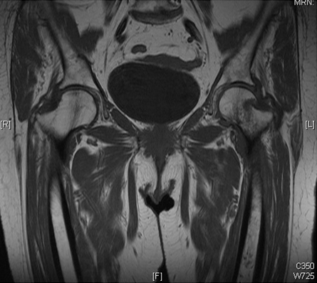
MRI showing coronal imaging confirming an intracapsular fracture of the left hip
From the collection of Bradley A. Petrisor, MSc, MD, FRCSC and Mohit Bhandari, MD, MSc, FRCSC
See this image in context in the following section/s:
Hip fractures
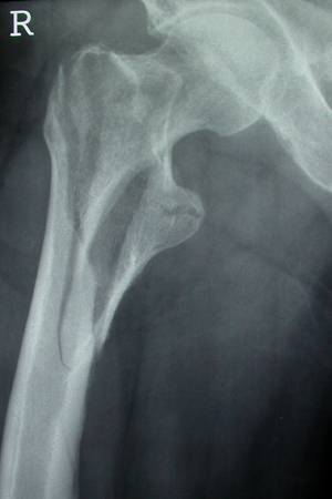
Unstable intertrochanteric fracture on x-ray
From the collection of Bradley A. Petrisor, MSc, MD, FRCSC and Mohit Bhandari, MD, MSc, FRCSC
See this image in context in the following section/s:

Hip fractures
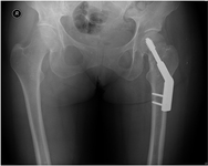
Anteroposterior pelvic radiograph showing a left intracapsular fracture fixed with a sliding hip screw construct
From the collection of Bradley A. Petrisor, MSc, MD, FRCSC and Mohit Bhandari, MD, MSc, FRCSC
See this image in context in the following section/s:
Hip fractures
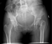
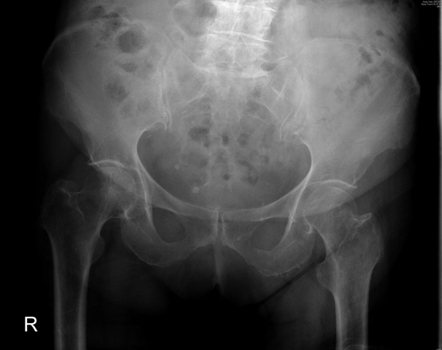
Anteroposterior pelvic radiograph showing a possible intracapsular fracture of the left hip
From the collection of Bradley A. Petrisor, MSc, MD, FRCSC and Mohit Bhandari, MD, MSc, FRCSC
See this image in context in the following section/s:
Hip fractures
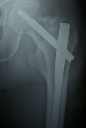
Intramedullary nail (cephalomedullary) for the treatment of an unstable intertrochanteric fracture
From the collection of Bradley A. Petrisor, MSc, MD, FRCSC and Mohit Bhandari, MD, MSc, FRCSC
See this image in context in the following section/s:
Hip fractures
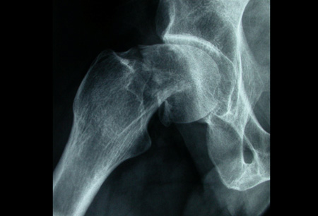
Anteroposterior x-ray showing femoral neck fracture
From the collection of Bradley A. Petrisor, MSc, MD, FRCSC and Mohit Bhandari, MD, MSc, FRCSC
See this image in context in the following section/s:

Hip fractures
Initial anteroposterior radiograph showing a displaced left hip intracapsular fracture
From the collection of Bradley A. Petrisor, MSc, MD, FRCSC and Mohit Bhandari, MD, MSc, FRCSC
See this image in context in the following section/s:
Use of this content is subject to our disclaimer