Images and videos
Images

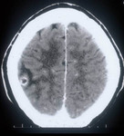

Tapeworm infection
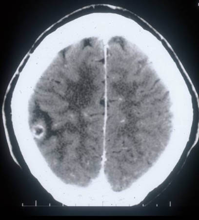
Colloidal stage - neurocysticercosis: CT scan showing ring enhancing cystic lesion in the temporal lobe and perilesional edema
From the personal collections of Dr Christina Coyle and Dr Maheen Saeed; used with permission
See this image in context in the following section/s:
Tapeworm infection
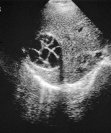
Echinococcus of liver with daughter cyst on ultrasound: multivesicular, multiseptated cysts, where daughter cyst completely fills the unilocular mother cyst; cyst produces a wheel-like structure
From the personal collections of Dr Christina Coyle and Dr Maheen Saeed; used with permission
See this image in context in the following section/s:

Tapeworm infection
Granular stage - neurocysticercosis: MRI scan showing enhancing lesion without perilesional edema
From the personal collections of Dr Christina Coyle and Dr Maheen Saeed; used with permission
See this image in context in the following section/s:

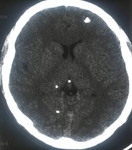
Tapeworm infection
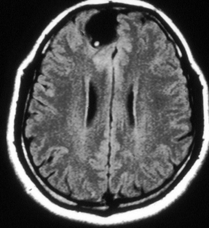
Calcified stage - neurocysticercosis: MRI scan of multiple calcified lesions in a patient with neurocysticercosis
From the personal collections of Dr Christina Coyle and Dr Maheen Saeed; used with permission
See this image in context in the following section/s:
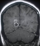
Tapeworm infection
Cystic stage - neurocysticercosis: MRI scan showing cystic lesion in the frontal lobe; a scolex can be seen within the cyst
From the personal collections of Dr Christina Coyle and Dr Maheen Saeed; used with permission
See this image in context in the following section/s:

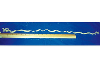
Tapeworm infection
Adult tapeworm identified as Taenia saginata
From the personal collections of Dr Christina Coyle and Dr Maheen Saeed; used with permission
See this image in context in the following section/s:
Use of this content is subject to our disclaimer