Etiology
AR can be caused by primary disease of the aortic valve leaflets or dilation of the aortic root. In developing countries rheumatic heart disease is the most common cause, but congenital bicuspid aortic valve and aortic root dilation account for most of the cases in developed countries.[1] Causes of aortic root dilation include Marfan syndrome, related connective tissue diseases, and aortitis secondary to syphilis, Behcet, Takayasu, reactive arthritis, or ankylosing spondylitis. Endocarditis can lead to rupture of leaflets or even paravalvular leaks. Vegetations on the valvular cusps can also cause inadequate closure of leaflets, resulting in leakage of blood.[7] Aortic root dissection is a cause of acute AR.
AR may develop acutely (acute AR) or over a period of many years in progressively increasing severity (chronic AR). An analysis of the causes of AR in patients >20 years of age undergoing isolated aortic valve replacement/repair (AVR) in a single US tertiary center from 1993-2005, found nonvalvular causes accounted for 54% of cases and valvular causes for 46%. Acute AR was responsible for only 18% of AVR, and, of these, 56% resulted from active infective endocarditis and 44% from aortic dissection. Aortic enlargement from unclear etiology was the most common cause of chronic AR, accounting for 34% of the total, followed by bicuspid congenital malformation (22% of total). Older patients most commonly had an unclear etiology.[8]
The role of systemic hypertension in aortic root dilation leading to AR is a frequent source of debate. Aortic root diameter at the supra-aortic ridge, which is the site of commissural attachment, is significantly greater in hypertensive patients than in age- and sex-matched normotensive patients.[9][Figure caption and citation for the preceding image starts]: Causes of ARDr Sanjeev Wasson and Dr Nishant Kalra; used with permission [Citation ends].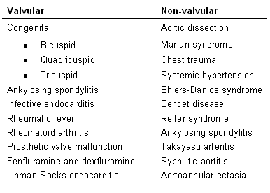
Pathophysiology
AR can present acutely or over decades.
Acute AR is a medical emergency with high mortality and results in an acute rise in left atrial pressure, pulmonary edema, and cardiogenic shock.
During acute AR:
End-diastolic pressure in the left ventricle rises sharply.
The heart tries to compensate by increasing the heart rate and increasing the contractility (Starling's law) to keep up with the increased preload, but this is insufficient to maintain the normal stroke volume and fails.
Chronic progressive AR results in:
Both left ventricular volume and pressure overload.
An increase in left ventricular volume and pressure causes an increase in wall tension.
According to Laplace's law, wall tension is directly proportional to the product of cavity pressure and radius, and inversely proportional to wall thickness.
To compensate for the increased wall tension, the heart wall undergoes hypertrophy. Both concentric and eccentric hypertrophy can occur but most are eccentric. Eccentric hypertrophy, in which sarcomeres are laid down in series, results from volume overload; concentric hypertrophy, in which sarcomeres replicate in parallel, results from pressure overload from increased systolic pressure to normalize the end-systolic stress.[10]
Systolic hypertension occurs secondary to increased stroke volume, which combines both regurgitant and forward stroke volume.
The volume overload, which is directly related to the severity of the leak, results in an increase in left ventricular end-diastolic volume.
End-diastolic pressure remains normal due to an increase in ventricular compliance resulting from increased cavity size.
In chronic AR, most patients remain asymptomatic for decades, as the left ventricle maintains forward stroke volume with compensatory chamber enlargement and hypertrophy. Eventually, the left ventricular systolic dysfunction supervenes and left ventricular end-diastolic pressure rises resulting in symptomatic congestive heart failure. Timing AVR before irreversible myocardial dysfunction develops is of critical importance.
Classification
Acute aortic regurgitation: clinically accepted criteria
A medical emergency where the left heart rapidly decompensates due to its inability to handle a sudden increase in end-diastolic volume. Most commonly it results from aortic dissection or endocarditis and, in rare cases, trauma.
Chronic aortic regurgitation: clinically accepted criteria
Chronic regurgitation has a prolonged course over a period of months to years. The left ventricle is able to compensate for volume overload initially but then decompensates with the appearance of clinical symptoms of congestive heart failure.
Use of this content is subject to our disclaimer