Images and videos
Images

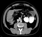

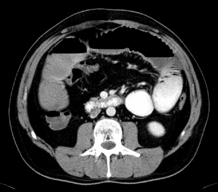
Ileus
CT scan with intravenous and oral contrast showing fluid-filled small intestine and cecum in ileus
From the personal collection of Dr Paula I. Denoya
See this image in context in the following section/s:

Ileus
CT scan with intravenous and oral contrast showing fluid-filled small intestine in ileus
From the personal collection of Dr Paula I. Denoya
See this image in context in the following section/s:

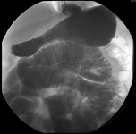
Ileus
CT scan showing significantly dilated stomach
From the personal collection of Dr Paula I. Denoya
See this image in context in the following section/s:

Ileus
Small bowel series showing dilated small bowel loops in ileus; nasogastric tube is seen curled in the stomach
From the personal collection of Dr Paula I. Denoya
See this image in context in the following section/s:

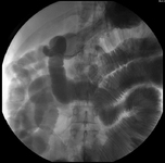
Ileus
Small bowel series showing dilated contrast-filled small bowel loops in ileus; some contrast is visible in the right colon
From the personal collection of Dr Paula I. Denoya
See this image in context in the following section/s:

Ileus
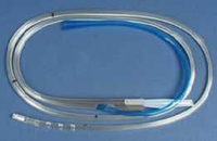
Nasogastric tube
From the personal collection of Dr Paula I. Denoya
See this image in context in the following section/s:
Videos
 Nasogastric tube insertion animated demonstration
How to insert a fine bore nasogastric tube for feeding.
Use of this content is subject to our disclaimer