Differentials
Vitiligo
SIGNS / SYMPTOMS
Lesions are not associated with scarring or architectural changes; typically asymptomatic, and do not cause pruritus or pain.[70][71]
Areas of vitiligo-affected skin are smooth, without textural changes associated with lichen sclerosus (e.g., lichenification/hyperkeratosis, atrophy).
[Figure caption and citation for the preceding image starts]: Vitiligo affecting the anus/perineumVitiligo Anus, Wikimedia Commons; public domain [Citation ends].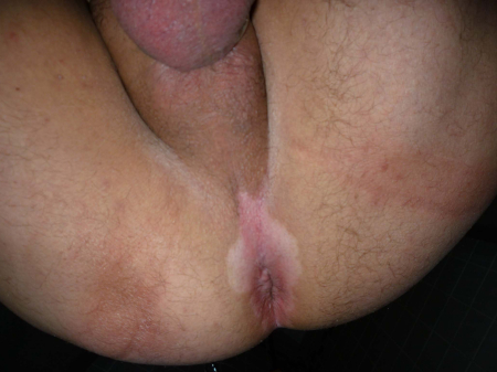
INVESTIGATIONS
Punch biopsy: Fontana-Masson stain on tissue that shows absence of melanin in vitiligo.
Wood lamp exam: can highlight the depigmentation in vitiligo as sharp borders that appear bright blue/white (note that LS can also show depigmentation in long-standing lesions so a Wood lamp exam is not specific for vitiligo).
Lichen planus
SIGNS / SYMPTOMS
Most commonly presents with erosions; Wickham’s striae (white lacy network overlying lesions) are typically present.
There may be oral or esophageal mucosal involvement; other cutaneous sites may also be involved (e.g., lichen planopilaris of the scalp, dorsal pterygium of the nails).[72] Rarely, ocular involvement may also be noted (also consider mucous membrane pemphigoid as a differential in these cases).
Lichen planus may affect the vagina, which is a very rare feature in LS unless significant vaginal prolapse is present.
[Figure caption and citation for the preceding image starts]: Active lichen planus - purplish-violaceous, polygonal-shaped, flat-topped small papules located on the forearms, with presence of Wickham’s striae and excoriationsLitaiem N et al. Case Reports 2016; used with permission [Citation ends].
INVESTIGATIONS
Punch biopsy of transition zone between normal skin and erosions: lichenoid band of inflammation with sawtooth rete ridge pattern.
Greater degree of lichenoid interface dermatitis (inflammation at junction of epidermis and dermis) in LP. Absence of sclerotic changes.
Immunochemistry with CD34: presence of CD34 in affected areas.
Candidiasis
SIGNS / SYMPTOMS
Typically bright red in color; may have satellite papules. No architectural changes, hypopigmentation, or depigmentation. Lesions are not atrophic.
May or may not have vaginal discharge/inflammation.[73]
Lichen simplex chronicus
SIGNS / SYMPTOMS
Commonly presents with skin thickening/lichenification involving one or both sides of the vulva. Excoriations and exaggerated skin lines may be seen. May involve the mucous membranes including the mouth and vagina.
Note: can be seen as an overlapping condition in LS.
INVESTIGATIONS
Standard histology shows compact orthokeratosis, psoriasiform acanthosis of the epidermis, vertically oriented dermal blood vessels, and collagen in the papillary dermis.
Irritant contact dermatitis
SIGNS / SYMPTOMS
No architectural changes; no hypopigmentation or depigmented atrophic plaques.[74]
[Figure caption and citation for the preceding image starts]: Allergic contact dermatitis to nickel in watchbandFrom the personal collection of Dr Snehal Desai; used with permission [Citation ends].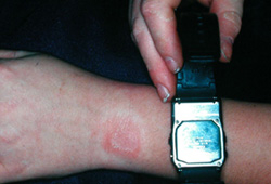
INVESTIGATIONS
Patch testing: may identify causative agent.
Genitourinary syndrome of menopause (GSM)
SIGNS / SYMPTOMS
Found in postmenopausal women or those taking suppressive oral contraceptives. Other symptoms of menopause, such as irregular menstrual cycle, heavy menstrual bleeding/amenorrhea, mood changes, hot flashes, and night sweats, may be present.
There may be pallor of vaginal mucosa, but no scarring or associated architectural changes.
Local estrogen therapy will result in resolution in symptoms.[75]
INVESTIGATIONS
Wet mount or vaginal exam can demonstrate atrophic changes of the vaginal mucosa; parabasal cells on wet mount and vaginal pallor on speculum exam.[45]
Vestibular sclerosis
Psoriasis
SIGNS / SYMPTOMS
Not associated with architectural changes.
Skin lesions appear as erythematous, well-demarcated or “glazed” plaques with or without scale, which usually involve extensor surfaces such as the elbows and knees. Lesions may also occur in the gluteal cleft or scalp. Patients may also have associated nail disease.
[Figure caption and citation for the preceding image starts]: Plaque psoriasis on kneeFrom the collection of Professor Tsu-Yi Chuang, MD, MPH, FAAD; used with permission [Citation ends].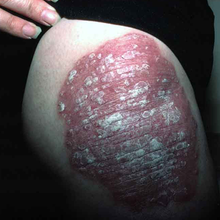
INVESTIGATIONS
Biopsy: histology will reveal psoriasiform epidermal hyperplasia and other classic features of psoriasis such as regular acanthosis, loss of the granular layer, thinning of the suprapapillary plates, dilation of blood vessels in the superficial papillary dermis, neutrophilic abscesses in the stratum corneum, and/or parakeratosis.[78] However, note that classic features of psoriasis found elsewhere often are absent in intertriginous psoriasis.
Biopsies of vulvar areas of psoriasis may also demonstrate spongiosis that is often absent in other clinical sites.[79]
Morphea
SIGNS / SYMPTOMS
Often more indurated and usually more commonly found on extragenital sites. Difficult to distinguish clinically on extragenital sites from LS.
Note that morphea and lichen sclerosus tend to overlap and can occur in the same patient.[69][80]
INVESTIGATIONS
Biopsy can provide clues to diagnosis as morphea tends to have perivascular infiltration and involvement of the reticular dermis with the destruction of the adnexal structures.[81]
Use of this content is subject to our disclaimer