Images and videos
Images


Evaluation of dysuria
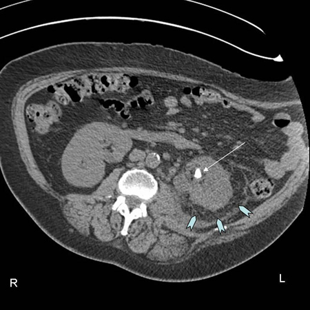
CT scan showing left renal stone (white arrow) with perinephric stranding around the left kidney (blue chevrons) and pyelonephritis
From the personal collection of Dr Kasra Saeb-Parsy
See this image in context in the following section/s:
Evaluation of dysuria
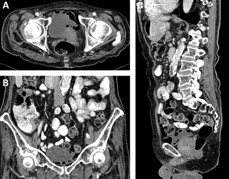
Emphysematous cystitis: (A) horizontal CT slice showing increased emphysema, (B) coronal CT slice showing increased emphysema, (C) sagittal CT slice showing increased emphysema
Middela S, Green E, Montague R. Emphysematous cystitis: radiological diagnosis of complicated urinary tract infection. BMJ Case Rep. 2009; doi:10.1136/bcr.05.2009.1832. Used with permission
See this image in context in the following section/s:
Evaluation of dysuria
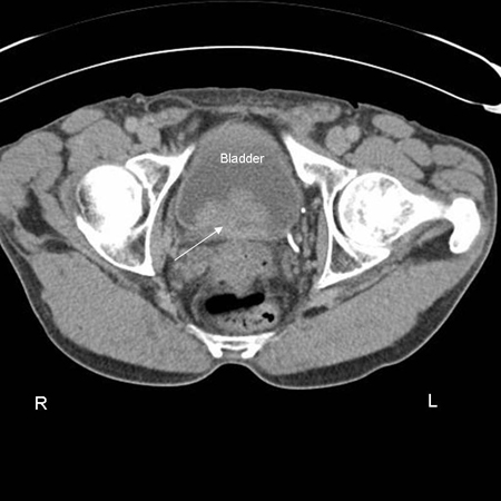
CT scan showing transitional cell carcinoma of the bladder blocking the right ureteric orifice
From the personal collection of Dr Kasra Saeb-Parsy
See this image in context in the following section/s:

Evaluation of dysuria
CT scan showing left ureteric calculi
From the personal collection of Dr Kasra Saeb-Parsy
See this image in context in the following section/s:
Use of this content is subject to our disclaimer