The main goal of treatment in sudden cardiac arrest is to achieve a return of spontaneous (as opposed to cardiopulmonary resuscitation [CPR]-mediated) circulation.[68]Desbiens NA. Simplifying the diagnosis and management of pulseless electrical activity in adults: a qualitative review. Crit Care Med. 2008;36:391-6.
http://www.ncbi.nlm.nih.gov/pubmed/18216597?tool=bestpractice.com
The algorithm of basic life support (BLS) and advanced cardiac life support (ACLS) provided by the American Heart Association (AHA) gives a systematic approach to the treatment of sudden cardiac arrest.[1]Panchal AR, Bartos JA, Cabañas JG, et al. Part 3: adult basic and advanced life support: 2020 American Heart Association guidelines for cardiopulmonary resuscitation and emergency cardiovascular care. Circulation. 2020 Oct 20;142(16_suppl_2):S366-468.
https://www.ahajournals.org/doi/full/10.1161/CIR.0000000000000916
http://www.ncbi.nlm.nih.gov/pubmed/33081529?tool=bestpractice.com
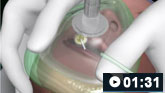
The methods vary slightly based on the underlying rhythm and cause of sudden cardiac arrest, but all rely on immediate attention to stabilizing the patient's respiratory status, addressing airway management as needed, and providing medications and other life-saving treatments aimed at correcting the unstable rhythm, as well as treating the underlying cause, all the while providing compressions to preserve vital organ perfusion.
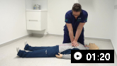
After activation of emergency medical services, the first approach to sudden cardiac arrest is BLS, providing compressions (first priority), assessing the airway, and giving breaths. This C-A-B priority recommendation by the AHA for adults is a change in the guidelines from the A-B-C priority taught historically, in order to emphasize the importance of providing timely chest compressions.[1]Panchal AR, Bartos JA, Cabañas JG, et al. Part 3: adult basic and advanced life support: 2020 American Heart Association guidelines for cardiopulmonary resuscitation and emergency cardiovascular care. Circulation. 2020 Oct 20;142(16_suppl_2):S366-468.
https://www.ahajournals.org/doi/full/10.1161/CIR.0000000000000916
http://www.ncbi.nlm.nih.gov/pubmed/33081529?tool=bestpractice.com
[69]Rea TD, Fahrenbruch C, Culley L, et al. CPR with chest compression alone or with rescue breathing. N Engl J Med. 2010;363:423-33.
http://www.ncbi.nlm.nih.gov/pubmed/20818863?tool=bestpractice.com
[70]Svensson L, Bohm K, Castrèn M, et al. Compression-only CPR or standard CPR in out-of-hospital cardiac arrest. N Engl J Med. 2010;363:434-42.
http://www.ncbi.nlm.nih.gov/pubmed/20818864?tool=bestpractice.com
Untrained lay-rescuers should perform compression-only CPR.[1]Panchal AR, Bartos JA, Cabañas JG, et al. Part 3: adult basic and advanced life support: 2020 American Heart Association guidelines for cardiopulmonary resuscitation and emergency cardiovascular care. Circulation. 2020 Oct 20;142(16_suppl_2):S366-468.
https://www.ahajournals.org/doi/full/10.1161/CIR.0000000000000916
http://www.ncbi.nlm.nih.gov/pubmed/33081529?tool=bestpractice.com
For rescuers trained in CPR using chest compressions and ventilation, it is reasonable to provide rescue breaths in addition to compressions.[1]Panchal AR, Bartos JA, Cabañas JG, et al. Part 3: adult basic and advanced life support: 2020 American Heart Association guidelines for cardiopulmonary resuscitation and emergency cardiovascular care. Circulation. 2020 Oct 20;142(16_suppl_2):S366-468.
https://www.ahajournals.org/doi/full/10.1161/CIR.0000000000000916
http://www.ncbi.nlm.nih.gov/pubmed/33081529?tool=bestpractice.com
[51]Wyckoff MH, Greif R, Morley PT, et al. 2022 International consensus on cardiopulmonary resuscitation and emergency cardiovascular care science with treatment recommendations: summary from the Basic Life Support; Advanced Life Support; Pediatric Life Support; Neonatal Life Support; Education, Implementation, and Teams; and First Aid task forces. Circulation. 3 Nov 2022 [Epub ahead of print].
http://www.ncbi.nlm.nih.gov/pubmed/36325905?tool=bestpractice.com
In cases where it is suspected that opioid overdose has caused the cardiac arrest, naloxone should be administered.[37]Dezfulian C, Orkin AM, Maron BA, et al. Opioid-associated out-of-hospital cardiac arrest: distinctive clinical features and implications for health care and public responses: a scientific statement from the American Heart Association. Circulation. 2021 Apr 20;143(16):e836-e870.
https://www.ahajournals.org/doi/reader/10.1161/CIR.0000000000000958
http://www.ncbi.nlm.nih.gov/pubmed/33682423?tool=bestpractice.com
Patients who require further treatment are then given ACLS by trained providers.[1]Panchal AR, Bartos JA, Cabañas JG, et al. Part 3: adult basic and advanced life support: 2020 American Heart Association guidelines for cardiopulmonary resuscitation and emergency cardiovascular care. Circulation. 2020 Oct 20;142(16_suppl_2):S366-468.
https://www.ahajournals.org/doi/full/10.1161/CIR.0000000000000916
http://www.ncbi.nlm.nih.gov/pubmed/33081529?tool=bestpractice.com
At any given point in the provision of ACLS, the rhythm may change from pulseless ventricular tachycardia (VT)/ventricular fibrillation (VF) to pulseless electrical activity (PEA)/asystole, or vice versa. In such an event, the appropriate ACLS algorithm for the new rhythm should be followed.
Epinephrine is recommended for both shockable and nonshockable rhythms.[1]Panchal AR, Bartos JA, Cabañas JG, et al. Part 3: adult basic and advanced life support: 2020 American Heart Association guidelines for cardiopulmonary resuscitation and emergency cardiovascular care. Circulation. 2020 Oct 20;142(16_suppl_2):S366-468.
https://www.ahajournals.org/doi/full/10.1161/CIR.0000000000000916
http://www.ncbi.nlm.nih.gov/pubmed/33081529?tool=bestpractice.com
[50]Soar J, Böttiger BW, Carli P, et al. European Resuscitation Council guidelines 2021: adult advanced life support. Resuscitation. 2021 Apr;161:115-51.
http://www.ncbi.nlm.nih.gov/pubmed/33773825?tool=bestpractice.com
[54]Perman SM, Elmer J, Maciel CB, et al. 2023 American Heart Association focused update on adult advanced cardiovascular life support: an update to the American Heart Association guidelines for cardiopulmonary resuscitation and emergency cardiovascular care. Circulation. 2024 Jan 30;149(5):e254-73.
https://www.ahajournals.org/doi/full/10.1161/CIR.0000000000001194
http://www.ncbi.nlm.nih.gov/pubmed/38108133?tool=bestpractice.com
The use of epinephrine during cardiac arrest has been shown to increase the rate of achieving return of spontaneous circulation (ROSC) and to increase short-term survival.[71]Perkins GD, Ji C, Achana F, et al. Adrenaline to improve survival in out-of-hospital cardiac arrest: the PARAMEDIC2 RCT. Health Technol Assess. 2021 Apr;25(25):1-166.
http://www.ncbi.nlm.nih.gov/pubmed/33861194?tool=bestpractice.com
[72]Perkins GD, Ji C, Deakin CD, et al. A randomized trial of epinephrine in out-of-hospital cardiac arrest. N Engl J Med. 2018 Aug 23;379(8):711-21.
https://www.nejm.org/doi/10.1056/NEJMoa1806842
http://www.ncbi.nlm.nih.gov/pubmed/30021076?tool=bestpractice.com
[73]Holmberg MJ, Issa MS, Moskowitz A, et al. Vasopressors during adult cardiac arrest: a systematic review and meta-analysis. Resuscitation. 2019 Jun;139:106-21.
https://www.resuscitationjournal.com/article/S0300-9572(19)30122-4/fulltext
http://www.ncbi.nlm.nih.gov/pubmed/30980877?tool=bestpractice.com
[74]Finn J, Jacobs I, Williams TA, et al. Adrenaline and vasopressin for cardiac arrest. Cochrane Database Syst Rev. 2019 Jan 17;(1):CD003179.
https://www.cochranelibrary.com/cdsr/doi/10.1002/14651858.CD003179.pub2/full
http://www.ncbi.nlm.nih.gov/pubmed/30653257?tool=bestpractice.com
However, epinephrine use during cardiac arrest has not been shown to lead to significantly improved neurologic outcomes, and may lead to higher rates of severe neurologic impairment among survivors.[71]Perkins GD, Ji C, Achana F, et al. Adrenaline to improve survival in out-of-hospital cardiac arrest: the PARAMEDIC2 RCT. Health Technol Assess. 2021 Apr;25(25):1-166.
http://www.ncbi.nlm.nih.gov/pubmed/33861194?tool=bestpractice.com
[72]Perkins GD, Ji C, Deakin CD, et al. A randomized trial of epinephrine in out-of-hospital cardiac arrest. N Engl J Med. 2018 Aug 23;379(8):711-21.
https://www.nejm.org/doi/10.1056/NEJMoa1806842
http://www.ncbi.nlm.nih.gov/pubmed/30021076?tool=bestpractice.com
[73]Holmberg MJ, Issa MS, Moskowitz A, et al. Vasopressors during adult cardiac arrest: a systematic review and meta-analysis. Resuscitation. 2019 Jun;139:106-21.
https://www.resuscitationjournal.com/article/S0300-9572(19)30122-4/fulltext
http://www.ncbi.nlm.nih.gov/pubmed/30980877?tool=bestpractice.com
[74]Finn J, Jacobs I, Williams TA, et al. Adrenaline and vasopressin for cardiac arrest. Cochrane Database Syst Rev. 2019 Jan 17;(1):CD003179.
https://www.cochranelibrary.com/cdsr/doi/10.1002/14651858.CD003179.pub2/full
http://www.ncbi.nlm.nih.gov/pubmed/30653257?tool=bestpractice.com
One large randomized controlled trial (PARAMEDIC2) found no significant difference in the proportion of patients surviving to discharge with favorable neurologic outcome.[71]Perkins GD, Ji C, Achana F, et al. Adrenaline to improve survival in out-of-hospital cardiac arrest: the PARAMEDIC2 RCT. Health Technol Assess. 2021 Apr;25(25):1-166.
http://www.ncbi.nlm.nih.gov/pubmed/33861194?tool=bestpractice.com
While performing ACLS, the care team will assess for and treat any suspected reversible causes of cardiac arrest. If poisoning is suspected or confirmed, timely consult with a toxicologist or regional poison center should be undertaken to facilitate rapid and effective therapy. Treatment of cardiac arrest and life-threatening toxicity due to poisoning often requires specialized treatments that most clinicians do not use frequently, such as antidotes and venoarterial extracorporeal membrane oxygenation, in addition to effective basic and advanced life support. Guidelines have been published for the specific management of cardiac arrest due to critical poisoning from benzodiazepines, beta-blockers, calcium channel blockers, cocaine, cyanide, digoxin and related cardiac glycosides, local anesthetics, methemoglobinemia, opioids, organophosphates and carbamates, sodium channel antagonists, and sympathomimetics.[35]Lavonas EJ, Akpunonu PD, Arens AM, et al. 2023 American Heart Association focused update on the management of patients with cardiac arrest or life-threatening toxicity due to poisoning: an update to the American Heart Association guidelines for cardiopulmonary resuscitation and emergency cardiovascular care. Circulation. 2023 Oct 17;148(16):e149-84.
https://www.ahajournals.org/doi/10.1161/CIR.0000000000001161
http://www.ncbi.nlm.nih.gov/pubmed/37721023?tool=bestpractice.com
US guidelines published during the COVID-19 pandemic advise that chest compressions or defibrillation should not be delayed for providers to don personal protective equipment (PPE), but that initial resuscitation personnel should be relieved by providers wearing appropriate PPE as soon as possible.[75]Hsu A, Sasson C, Kudenchuk PJ, et al. 2021 Interim guidance to health care providers for basic and advanced cardiac life support in adults, children, and neonates with suspected or confirmed COVID-19. Circ Cardiovasc Qual Outcomes. 2021 Oct;14(10):e008396.
https://www.ahajournals.org/doi/10.1161/CIRCOUTCOMES.121.008396
If the patient cannot be placed supine, cardiopulmonary resuscitation may be provided in the prone position, particularly if the patient has advanced airway and circulatory support.[76]Task Force for the management of COVID-19 of the European Society of Cardiology. ESC guidance for the diagnosis and management of cardiovascular disease during the COVID-19 pandemic: part 2-care pathways, treatment, and follow-up. Eur Heart J. 2022 Mar 14;43(11):1059-103.
https://academic.oup.com/eurheartj/article/43/11/1059/6429145
http://www.ncbi.nlm.nih.gov/pubmed/34791154?tool=bestpractice.com
UK guidance advises that for those working in healthcare settings, the use of FFP3 masks or respirators and eye protection is recommended when performing chest compressions for patients with suspected or confirmed COVID-19. PPE should be donned as swiftly as possible to avoid any delays in treatment.[77]Resuscitation Council UK. Guidance: COVID-19: update to Resuscitation Council UK (RCUK) guidance for practice. Aug 2022 [internet publication].
https://www.resus.org.uk/library/additional-guidance/guidance-covid-19
Shockable rhythms (pulseless VT and VF)
In the setting of pulseless VT/VF, the initial management is of BLS as described above (C-A-B method). Early provision of CPR, including compression-only CPR, by bystanders in out-of-hospital arrest increases the rate of survival.[7]Zeppenfeld K, Tfelt-Hansen J, de Riva M, et al. 2022 ESC Guidelines for the management of patients with ventricular arrhythmias and the prevention of sudden cardiac death. Eur Heart J. 2022 Oct 21;43(40):3997-4126.
https://academic.oup.com/eurheartj/article/43/40/3997/6675633?login=false
http://www.ncbi.nlm.nih.gov/pubmed/36017572?tool=bestpractice.com
[78]Gallagher EJ, Lombardi G, Gennis P. Effectiveness of bystander cardiopulmonary resuscitation and survival following out-of-hospital cardiac arrest. JAMA. 1995;274:1922-5.
http://www.ncbi.nlm.nih.gov/pubmed/8568985?tool=bestpractice.com
[79]Zhan L, Yang LJ, Huang Y, et al. Continuous chest compression versus interrupted chest compression for cardiopulmonary resuscitation of non-asphyxial out-of-hospital cardiac arrest. Cochrane Database Syst Rev. 2017;(3):CD010134.
http://onlinelibrary.wiley.com/doi/10.1002/14651858.CD010134.pub2/abstract
http://www.ncbi.nlm.nih.gov/pubmed/28349529?tool=bestpractice.com
Laypeople in the US initiated CPR in 40% of out-of-hospital cardiac arrests (OHCA) in 2022.[2]Martin SS, Aday AW, Almarzooq ZI, et al. 2024 heart disease and stroke statistics: a report of US and global data from the American Heart Association. Circulation. 2024 Feb 20;149(8):e347-913.
https://www.doi.org/10.1161/CIR.0000000000001209
http://www.ncbi.nlm.nih.gov/pubmed/38264914?tool=bestpractice.com
Work has shown that compression-only resuscitation by bystanders for OHCA is equally, if not more, efficient in providing life-saving therapy, compared with conventional CPR with rescue breaths.[1]Panchal AR, Bartos JA, Cabañas JG, et al. Part 3: adult basic and advanced life support: 2020 American Heart Association guidelines for cardiopulmonary resuscitation and emergency cardiovascular care. Circulation. 2020 Oct 20;142(16_suppl_2):S366-468.
https://www.ahajournals.org/doi/full/10.1161/CIR.0000000000000916
http://www.ncbi.nlm.nih.gov/pubmed/33081529?tool=bestpractice.com
[80]SOS-KANTO study group. Cardiopulmonary resuscitation by bystanders with chest compression only (SOS-KANTO): an observational study. Lancet. 2007;369:920-6.
http://www.ncbi.nlm.nih.gov/pubmed/17368153?tool=bestpractice.com
In a survey of 9022 people in the US in 2015, the prevalence of reported current training in CPR was 18%, and the prevalence of having CPR training at some point was 65%. The rates were lower in Hispanic/Latino people, older people, people with less formal education, and lower-income groups.[81]Blewer AL, Ibrahim SA, Leary M, et al. Cardiopulmonary resuscitation training disparities in the United States. J Am Heart Assoc. 2017 May 17;6(5):e006124.
https://www.ahajournals.org/doi/10.1161/JAHA.117.006124
http://www.ncbi.nlm.nih.gov/pubmed/28515114?tool=bestpractice.com
Research supports increasing the availability of public-access defibrillators and community training in BLS methods, including training for schoolchildren.[7]Zeppenfeld K, Tfelt-Hansen J, de Riva M, et al. 2022 ESC Guidelines for the management of patients with ventricular arrhythmias and the prevention of sudden cardiac death. Eur Heart J. 2022 Oct 21;43(40):3997-4126.
https://academic.oup.com/eurheartj/article/43/40/3997/6675633?login=false
http://www.ncbi.nlm.nih.gov/pubmed/36017572?tool=bestpractice.com
[82]Schroeder DC, Semeraro F, Greif R, et al. KIDS SAVE LIVES: basic life support education for schoolchildren: a narrative review and scientific statement from the International Liaison Committee on Resuscitation. Circulation. 2023 Jun 13;147(24):1854-68.
https://www.ahajournals.org/doi/10.1161/CIR.0000000000001128
http://www.ncbi.nlm.nih.gov/pubmed/37194575?tool=bestpractice.com
BLS training should be particularly encouraged for likely rescuers of people at high risk of OHCA, such as those with cardiac disease, pulmonary disease, and drug-use disorder.[51]Wyckoff MH, Greif R, Morley PT, et al. 2022 International consensus on cardiopulmonary resuscitation and emergency cardiovascular care science with treatment recommendations: summary from the Basic Life Support; Advanced Life Support; Pediatric Life Support; Neonatal Life Support; Education, Implementation, and Teams; and First Aid task forces. Circulation. 3 Nov 2022 [Epub ahead of print].
http://www.ncbi.nlm.nih.gov/pubmed/36325905?tool=bestpractice.com
In Sweden, bystander CPR increased from 30.9% to 82.2% between 1990 and 2020, likely due to a 40-year campaign to educate the population about CPR.[83]Jerkeman M, Sultanian P, Lundgren P, et al. Trends in survival after cardiac arrest: a Swedish nationwide study over 30 years. Eur Heart J. 2022 Dec 7;43(46):4817-29.
https://academic.oup.com/eurheartj/article/43/46/4817/6655575
http://www.ncbi.nlm.nih.gov/pubmed/35924401?tool=bestpractice.com
ACLS is commenced when trained providers arrive. If spontaneous circulation is not restored and a shockable rhythm is identified, one shock should be delivered (120-200 J for biphasic or 360 J for monophasic) followed by 5 cycles (2 minutes) of CPR.[1]Panchal AR, Bartos JA, Cabañas JG, et al. Part 3: adult basic and advanced life support: 2020 American Heart Association guidelines for cardiopulmonary resuscitation and emergency cardiovascular care. Circulation. 2020 Oct 20;142(16_suppl_2):S366-468.
https://www.ahajournals.org/doi/full/10.1161/CIR.0000000000000916
http://www.ncbi.nlm.nih.gov/pubmed/33081529?tool=bestpractice.com
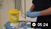
Intravenous (preferred) or intraosseous access is obtained without interrupting CPR.[1]Panchal AR, Bartos JA, Cabañas JG, et al. Part 3: adult basic and advanced life support: 2020 American Heart Association guidelines for cardiopulmonary resuscitation and emergency cardiovascular care. Circulation. 2020 Oct 20;142(16_suppl_2):S366-468.
https://www.ahajournals.org/doi/full/10.1161/CIR.0000000000000916
http://www.ncbi.nlm.nih.gov/pubmed/33081529?tool=bestpractice.com
The pulse and rhythm are again assessed and, if the patient remains in pulseless VT/VF, another equivalent or higher-energy shock is delivered and epinephrine given, followed by 5 cycles (2 minutes) of CPR.[1]Panchal AR, Bartos JA, Cabañas JG, et al. Part 3: adult basic and advanced life support: 2020 American Heart Association guidelines for cardiopulmonary resuscitation and emergency cardiovascular care. Circulation. 2020 Oct 20;142(16_suppl_2):S366-468.
https://www.ahajournals.org/doi/full/10.1161/CIR.0000000000000916
http://www.ncbi.nlm.nih.gov/pubmed/33081529?tool=bestpractice.com
[54]Perman SM, Elmer J, Maciel CB, et al. 2023 American Heart Association focused update on adult advanced cardiovascular life support: an update to the American Heart Association guidelines for cardiopulmonary resuscitation and emergency cardiovascular care. Circulation. 2024 Jan 30;149(5):e254-73.
https://www.ahajournals.org/doi/full/10.1161/CIR.0000000000001194
http://www.ncbi.nlm.nih.gov/pubmed/38108133?tool=bestpractice.com
If the rhythm is still pulseless VT/VF, another shock is delivered along with amiodarone
[  ]
How does amiodarone compare with other antiarrhythmic drugs for the secondary prevention of sudden cardiac death in at-risk adults?/cca.html?targetUrl=https://cochranelibrary.com/cca/doi/10.1002/cca.1276/fullShow me the answer or lidocaine, and CPR is continued for 5 cycles (2 minutes). If the patient remains in a shockable rhythm, the algorithm restarts at the stage of epinephrine administration. This cycle continues until spontaneous circulation is achieved or resuscitative measures are terminated.
]
How does amiodarone compare with other antiarrhythmic drugs for the secondary prevention of sudden cardiac death in at-risk adults?/cca.html?targetUrl=https://cochranelibrary.com/cca/doi/10.1002/cca.1276/fullShow me the answer or lidocaine, and CPR is continued for 5 cycles (2 minutes). If the patient remains in a shockable rhythm, the algorithm restarts at the stage of epinephrine administration. This cycle continues until spontaneous circulation is achieved or resuscitative measures are terminated.
In situations of witnessed arrest, electrical defibrillation should be attempted as soon as possible, not necessarily after 5 cycles (2 minutes) of CPR.[1]Panchal AR, Bartos JA, Cabañas JG, et al. Part 3: adult basic and advanced life support: 2020 American Heart Association guidelines for cardiopulmonary resuscitation and emergency cardiovascular care. Circulation. 2020 Oct 20;142(16_suppl_2):S366-468.
https://www.ahajournals.org/doi/full/10.1161/CIR.0000000000000916
http://www.ncbi.nlm.nih.gov/pubmed/33081529?tool=bestpractice.com
Due to the importance of prompt defibrillation, the use of "public access defibrillation" by lay-rescuers using automatic external defibrillators has gained favor and been found to increase the rate of sudden cardiac arrest patients surviving to hospital discharge.[7]Zeppenfeld K, Tfelt-Hansen J, de Riva M, et al. 2022 ESC Guidelines for the management of patients with ventricular arrhythmias and the prevention of sudden cardiac death. Eur Heart J. 2022 Oct 21;43(40):3997-4126.
https://academic.oup.com/eurheartj/article/43/40/3997/6675633?login=false
http://www.ncbi.nlm.nih.gov/pubmed/36017572?tool=bestpractice.com
[85]Tzivoni D, Banai S, Schuger C, et al. Treatment of torsade de pointes with magnesium sulfate. Circulation.1988; 77:392-7.
http://www.ncbi.nlm.nih.gov/pubmed/3338130?tool=bestpractice.com
[86]Sanna T, La Torre G, de Waure C, et al. Cardiopulmonary resuscitation alone vs. cardiopulmonary resuscitation plus automated external defibrillator use by non-healthcare professionals: a meta-analysis on 1583 cases of out-of-hospital cardiac arrest. Resuscitation. 2008;76:226-32.
http://www.ncbi.nlm.nih.gov/pubmed/17875357?tool=bestpractice.com
Lay-rescuer public access defibrillation has been shown to have a higher impact on survival than defibrillation by emergency dispatched professional first responders.[87]Bækgaard JS, Viereck S, Møller TP, et al. The effects of public access defibrillation on survival after out-of-hospital cardiac arrest: a systematic review of observational studies. Circulation. 2017 Sep 5;136(10):954-65.
https://www.ahajournals.org/doi/full/10.1161/CIRCULATIONAHA.117.029067
http://www.ncbi.nlm.nih.gov/pubmed/28687709?tool=bestpractice.com
The International Liaison Committee on Resuscitation suggest that use of a double sequential defibrillation strategy or vector change defibrillation strategy may be considered for adults who remain in pulseless VT/VF after 3 or more consecutive shocks (this is a weak recommendation, based on very low certainty evidence).[88]Greif R, Bray JE, Djärv T, et al. 2024 International consensus on cardiopulmonary resuscitation and emergency cardiovascular care science with treatment recommendations: summary from the basic life support; advanced life support; pediatric life support; neonatal life support; education, implementation, and teams; and first aid task forces. Resuscitation. 2024 Dec;205:110414.
https://www.resuscitationjournal.com/article/S0300-9572(24)00308-3/fulltext
http://www.ncbi.nlm.nih.gov/pubmed/39549953?tool=bestpractice.com
If a double sequential defibrillation strategy is used, it is good practice for one single operator to activate the defibrillators in sequence.[88]Greif R, Bray JE, Djärv T, et al. 2024 International consensus on cardiopulmonary resuscitation and emergency cardiovascular care science with treatment recommendations: summary from the basic life support; advanced life support; pediatric life support; neonatal life support; education, implementation, and teams; and first aid task forces. Resuscitation. 2024 Dec;205:110414.
https://www.resuscitationjournal.com/article/S0300-9572(24)00308-3/fulltext
http://www.ncbi.nlm.nih.gov/pubmed/39549953?tool=bestpractice.com
In patients with sudden cardiac arrest due to torsades de pointes, giving magnesium may restore a perfusing cardiac rhythm.[85]Tzivoni D, Banai S, Schuger C, et al. Treatment of torsade de pointes with magnesium sulfate. Circulation.1988; 77:392-7.
http://www.ncbi.nlm.nih.gov/pubmed/3338130?tool=bestpractice.com
[89]Reis AG, Ferreira de Paiva E, Schvartsman C, et al. Magnesium in cardiopulmonary resuscitation: critical review. Resuscitation. 2008;77:21-5.
http://www.ncbi.nlm.nih.gov/pubmed/18037222?tool=bestpractice.com
Nonshockable rhythms (PEA and asystole)
In the setting of PEA/asystole, the initial provision is of BLS as described above (C-A-B method).
ACLS is commenced when trained providers arrive. If spontaneous circulation is not restored and a nonshockable rhythm is identified, 5 cycles (2 minutes) of CPR are provided.[1]Panchal AR, Bartos JA, Cabañas JG, et al. Part 3: adult basic and advanced life support: 2020 American Heart Association guidelines for cardiopulmonary resuscitation and emergency cardiovascular care. Circulation. 2020 Oct 20;142(16_suppl_2):S366-468.
https://www.ahajournals.org/doi/full/10.1161/CIR.0000000000000916
http://www.ncbi.nlm.nih.gov/pubmed/33081529?tool=bestpractice.com
Intravenous (preferred) or intraosseous access is obtained without interrupting CPR, and epinephrine (adrenaline) is given as soon as possible and every 3-5 minutes thereafter. The pulse and rhythm are assessed after every 5 cycles (2 minutes) of CPR, and if a pulseless nonshockable rhythm remains, CPR is continued.[1]Panchal AR, Bartos JA, Cabañas JG, et al. Part 3: adult basic and advanced life support: 2020 American Heart Association guidelines for cardiopulmonary resuscitation and emergency cardiovascular care. Circulation. 2020 Oct 20;142(16_suppl_2):S366-468.
https://www.ahajournals.org/doi/full/10.1161/CIR.0000000000000916
http://www.ncbi.nlm.nih.gov/pubmed/33081529?tool=bestpractice.com
This cycle of giving CPR and epinephrine continues until spontaneous circulation is attained or resuscitation is terminated. In addition, empiric treatment for likely reversible causes may be considered, such as calcium bicarbonate for hyperkalemia in patients with a history of renal failure. There is no evidence to suggest that transcutaneous pacing should be used in patients with asystolic arrest.[90]Sherbino J, Verbeek PR, MacDonald RD, et al. Prehospital transcutaneous cardiac pacing for symptomatic bradycardia or bradyasystolic cardiac arrest: a systematic review. Resuscitation. 2006;70:193-200.
http://www.ncbi.nlm.nih.gov/pubmed/16814446?tool=bestpractice.com
Postresuscitation care
If ROSC is achieved, postresuscitation care should be instigated immediately. This involves continued monitoring, organ support, correction of electrolyte imbalances and acidosis, and safe transfer to a critical care environment. A thorough search for potential etiologies should be conducted, and risk factors for sudden cardiac arrest should be modified or treated.
A 12-lead ECG is recommended immediately after ROSC to determine whether signs of ST-elevation myocardial infarction (STEMI) are present.[1]Panchal AR, Bartos JA, Cabañas JG, et al. Part 3: adult basic and advanced life support: 2020 American Heart Association guidelines for cardiopulmonary resuscitation and emergency cardiovascular care. Circulation. 2020 Oct 20;142(16_suppl_2):S366-468.
https://www.ahajournals.org/doi/full/10.1161/CIR.0000000000000916
http://www.ncbi.nlm.nih.gov/pubmed/33081529?tool=bestpractice.com
In patients with STEMI, emergency coronary angiography, with or without percutaneous coronary intervention, should be performed.[54]Perman SM, Elmer J, Maciel CB, et al. 2023 American Heart Association focused update on adult advanced cardiovascular life support: an update to the American Heart Association guidelines for cardiopulmonary resuscitation and emergency cardiovascular care. Circulation. 2024 Jan 30;149(5):e254-73.
https://www.ahajournals.org/doi/full/10.1161/CIR.0000000000001194
http://www.ncbi.nlm.nih.gov/pubmed/38108133?tool=bestpractice.com
Emergency coronary angiography is also reasonable for select patients with suspected acute coronary syndrome without ST elevation, including those with hemodynamic/electrical instability or signs of ongoing ischemia.[54]Perman SM, Elmer J, Maciel CB, et al. 2023 American Heart Association focused update on adult advanced cardiovascular life support: an update to the American Heart Association guidelines for cardiopulmonary resuscitation and emergency cardiovascular care. Circulation. 2024 Jan 30;149(5):e254-73.
https://www.ahajournals.org/doi/full/10.1161/CIR.0000000000001194
http://www.ncbi.nlm.nih.gov/pubmed/38108133?tool=bestpractice.com
It is not recommended over delayed angiography in patients with ROSC in the absence of ST elevation, shock, electrical instability, signs of significant myocardial damage, or ongoing ischemia.[54]Perman SM, Elmer J, Maciel CB, et al. 2023 American Heart Association focused update on adult advanced cardiovascular life support: an update to the American Heart Association guidelines for cardiopulmonary resuscitation and emergency cardiovascular care. Circulation. 2024 Jan 30;149(5):e254-73.
https://www.ahajournals.org/doi/full/10.1161/CIR.0000000000001194
http://www.ncbi.nlm.nih.gov/pubmed/38108133?tool=bestpractice.com
Anoxic brain injury is a frequent complication of sudden cardiac arrest. A systematic review of the literature demonstrates that targeted temperature management (TTM) protocols improve survival and neurologic outcome following resuscitation from sudden cardiac arrest, with guidelines continuing to support their use.[1]Panchal AR, Bartos JA, Cabañas JG, et al. Part 3: adult basic and advanced life support: 2020 American Heart Association guidelines for cardiopulmonary resuscitation and emergency cardiovascular care. Circulation. 2020 Oct 20;142(16_suppl_2):S366-468.
https://www.ahajournals.org/doi/full/10.1161/CIR.0000000000000916
http://www.ncbi.nlm.nih.gov/pubmed/33081529?tool=bestpractice.com
[91]Donnino MW, Andersen LW, Berg KM, et al; ILCOR ALS Task Force. Temperature management after cardiac arrest: an advisory statement by the Advanced Life Support Task Force of the International Liaison Committee on Resuscitation and the American Heart Association Emergency Cardiovascular Care Committee and the Council on Cardiopulmonary, Critical Care, Perioperative and Resuscitation. Circulation. 2015 Dec 22;132(25):2448-56.
https://www.ahajournals.org/doi/full/10.1161/CIR.0000000000000313
http://www.ncbi.nlm.nih.gov/pubmed/26434495?tool=bestpractice.com
The American Heart Association (AHA) recommends that all patients unable to follow commands (i.e., are comatose) receive treatment that includes temperature control, regardless of their arrest location or presenting rhythm.[54]Perman SM, Elmer J, Maciel CB, et al. 2023 American Heart Association focused update on adult advanced cardiovascular life support: an update to the American Heart Association guidelines for cardiopulmonary resuscitation and emergency cardiovascular care. Circulation. 2024 Jan 30;149(5):e254-73.
https://www.ahajournals.org/doi/full/10.1161/CIR.0000000000001194
http://www.ncbi.nlm.nih.gov/pubmed/38108133?tool=bestpractice.com
There is a range for the target temperature, with more recent evidence suggesting that maintaining normothermia (i.e., avoidance of fever) may be equivalent to targeting hypothermia. One large randomized controlled trial (TTM2) which studied patients with coma after OHCA found no difference in 6 month survival or neurologic outcome in patients treated with hypothermia (target temperature of 91.4°F [33°C]) compared with normothermia (target temperature ≤99.5°F [≤37.5°C]).[92]Dankiewicz J, Cronberg T, Lilja G, et al. Hypothermia versus normothermia after out-of-hospital cardiac arrest. N Engl J Med. 2021 Jun 17;384(24):2283-94.
https://www.nejm.org/doi/10.1056/NEJMoa2100591?url_ver=Z39.88-2003&rfr_id=ori:rid:crossref.org&rfr_dat=cr_pub%20%200pubmed
http://www.ncbi.nlm.nih.gov/pubmed/34133859?tool=bestpractice.com
An earlier trial (TTM) found that a targeted temperature of 91.4°F (33°C) conferred no benefit compared with 96.8°F (36°C).[93]Nielsen N, Wetterslev J, Cronberg T, et al; TTM Trial Investigators. Targeted temperature management at 33°C versus 36°C after cardiac arrest. N Engl J Med. 2013;369:2197-206.
http://www.nejm.org/doi/full/10.1056/NEJMoa1310519
http://www.ncbi.nlm.nih.gov/pubmed/24237006?tool=bestpractice.com
For comatose adult patients with ROSC, AHA guidelines recommend targeting a temperature between 89.6°F and 99.5°F (32°C and 37.5°C) for at least 24 hours, and avoiding fever after the initial temperature control phase.[54]Perman SM, Elmer J, Maciel CB, et al. 2023 American Heart Association focused update on adult advanced cardiovascular life support: an update to the American Heart Association guidelines for cardiopulmonary resuscitation and emergency cardiovascular care. Circulation. 2024 Jan 30;149(5):e254-73.
https://www.ahajournals.org/doi/full/10.1161/CIR.0000000000001194
http://www.ncbi.nlm.nih.gov/pubmed/38108133?tool=bestpractice.com
[94]Perman SM, Bartos JA, Del Rios M, et al. Temperature management for comatose adult survivors of cardiac arrest: a science advisory from the American Heart Association. Circulation. 2023 Sep 19;148(12):982-8.
https://www.ahajournals.org/doi/10.1161/CIR.0000000000001164
http://www.ncbi.nlm.nih.gov/pubmed/37584195?tool=bestpractice.com
The 2024 International Liaison Committee on Resuscitation guidelines recommend actively preventing fever by targeting a temperature of ≤99.5°F (≤37.5°C) for 36-72 hours, commenting that the benefits of targeting hypothermia between 89.6°F and 93.2°F (32°C and 34°C) in selected subpopulations of patients remain uncertain.[88]Greif R, Bray JE, Djärv T, et al. 2024 International consensus on cardiopulmonary resuscitation and emergency cardiovascular care science with treatment recommendations: summary from the basic life support; advanced life support; pediatric life support; neonatal life support; education, implementation, and teams; and first aid task forces. Resuscitation. 2024 Dec;205:110414.
https://www.resuscitationjournal.com/article/S0300-9572(24)00308-3/fulltext
http://www.ncbi.nlm.nih.gov/pubmed/39549953?tool=bestpractice.com
European guidelines recommend targeting a temperature between 89.6°F and 96.8°F (32°C and 36°C).[95]Nolan JP, Sandroni C, Böttiger BW, et al. European Resuscitation Council and European Society of Intensive Care Medicine guidelines 2021: post-resuscitation care. Intensive Care Med. 2021 Apr;47(4):369-421.
https://link.springer.com/article/10.1007/s00134-021-06368-4
http://www.ncbi.nlm.nih.gov/pubmed/33765189?tool=bestpractice.com
TTM has three phases: induction, maintenance, and rewarming. Induction and/or maintenance can be achieved by:[95]Nolan JP, Sandroni C, Böttiger BW, et al. European Resuscitation Council and European Society of Intensive Care Medicine guidelines 2021: post-resuscitation care. Intensive Care Med. 2021 Apr;47(4):369-421.
https://link.springer.com/article/10.1007/s00134-021-06368-4
http://www.ncbi.nlm.nih.gov/pubmed/33765189?tool=bestpractice.com
Simple ice packs with or without wet towels
Cooling blankets or pads
Water- or air-circulating blankets
Water-circulating gel-coated pads
Transnasal evaporative cooling
Intravascular heat exchanger
Extracorporeal circulation.
Routine prehospital cooling of patients after ROSC with rapid infusion of cold intravenous fluids is not recommended.[1]Panchal AR, Bartos JA, Cabañas JG, et al. Part 3: adult basic and advanced life support: 2020 American Heart Association guidelines for cardiopulmonary resuscitation and emergency cardiovascular care. Circulation. 2020 Oct 20;142(16_suppl_2):S366-468.
https://www.ahajournals.org/doi/full/10.1161/CIR.0000000000000916
http://www.ncbi.nlm.nih.gov/pubmed/33081529?tool=bestpractice.com
[51]Wyckoff MH, Greif R, Morley PT, et al. 2022 International consensus on cardiopulmonary resuscitation and emergency cardiovascular care science with treatment recommendations: summary from the Basic Life Support; Advanced Life Support; Pediatric Life Support; Neonatal Life Support; Education, Implementation, and Teams; and First Aid task forces. Circulation. 3 Nov 2022 [Epub ahead of print].
http://www.ncbi.nlm.nih.gov/pubmed/36325905?tool=bestpractice.com
[95]Nolan JP, Sandroni C, Böttiger BW, et al. European Resuscitation Council and European Society of Intensive Care Medicine guidelines 2021: post-resuscitation care. Intensive Care Med. 2021 Apr;47(4):369-421.
https://link.springer.com/article/10.1007/s00134-021-06368-4
http://www.ncbi.nlm.nih.gov/pubmed/33765189?tool=bestpractice.com
Rewarming should be achieved slowly (0.45°F to 0.90°F [0.25°C to 0.50°C] of rewarming per hour) to avoid rebound hyperthermia, which is associated with worse neurologic outcomes.[95]Nolan JP, Sandroni C, Böttiger BW, et al. European Resuscitation Council and European Society of Intensive Care Medicine guidelines 2021: post-resuscitation care. Intensive Care Med. 2021 Apr;47(4):369-421.
https://link.springer.com/article/10.1007/s00134-021-06368-4
http://www.ncbi.nlm.nih.gov/pubmed/33765189?tool=bestpractice.com
There is evidence that patients who receive postresuscitation care at specialized centers have higher rates of neurologically intact survival, suggesting that postresuscitative treatment should ideally be performed in this setting.[96]Bosson N, Kaji AH, Niemann JT, et al. Survival and neurologic outcome after out-of-hospital cardiac arrest: results one year after regionalization of post-cardiac arrest care in a large metropolitan area. Prehosp Emerg Care. 2014 Apr-Jun;18(2):217-23.
http://www.ncbi.nlm.nih.gov/pubmed/24401209?tool=bestpractice.com
[97]Sinning C, Ahrens I, Cariou A, et al. The cardiac arrest centre for the treatment of sudden cardiac arrest due to presumed cardiac cause - aims, function and structure: Position paper of the Association for Acute CardioVascular Care of the European Society of Cardiology (AVCV), European Association of Percutaneous Coronary Interventions (EAPCI), European Heart Rhythm Association (EHRA), European Resuscitation Council (ERC), European Society for Emergency Medicine (EUSEM) and European Society of Intensive Care Medicine (ESICM). Eur Heart J Acute Cardiovasc Care. 2020 Nov;9(4_suppl):S193-S202.
https://academic.oup.com/ehjacc/article/9/4_suppl/S193/6125627?login=false
http://www.ncbi.nlm.nih.gov/pubmed/33327761?tool=bestpractice.com
Cardiac arrest centers have been shown to display higher coherence with guidelines compared with noncardiac arrest centers.[98]Jorge-Perez P, Nikolaou N, Donadello K, et al. Management of comatose survivors of out-of-hospital cardiac arrest in Europe: current treatment practice and adherence to guidelines. A joint survey by the Association for Acute CardioVascular Care (ACVC) of the ESC, the European Resuscitation Council (ERC), the European Society for Emergency Medicine (EUSEM), and the European Society of Intensive Care Medicine (ESICM). Eur Heart J Acute Cardiovasc Care. 2023 Feb 9;12(2):96-105.
https://academic.oup.com/ehjacc/article/12/2/96/6862066
http://www.ncbi.nlm.nih.gov/pubmed/36454812?tool=bestpractice.com
In-patient neurologic rehabilitation may be helpful for survivors who have suffered hypoxic-ischemic brain injury, although specific guidelines and evidence are lacking in this patient population.[95]Nolan JP, Sandroni C, Böttiger BW, et al. European Resuscitation Council and European Society of Intensive Care Medicine guidelines 2021: post-resuscitation care. Intensive Care Med. 2021 Apr;47(4):369-421.
https://link.springer.com/article/10.1007/s00134-021-06368-4
http://www.ncbi.nlm.nih.gov/pubmed/33765189?tool=bestpractice.com
Many patients will also be eligible for cardiac rehabilitation programs, which have been shown to reduce cardiovascular mortality and hospital admissions, and improve quality of life. They are mostly generic programs, in which patients with different cardiac diseases, for example, post acute coronary syndrome, heart failure, or post-cardiac surgery, can participate. They involve exercise training, risk factor management, lifestyle advice, education, and psychological support.[95]Nolan JP, Sandroni C, Böttiger BW, et al. European Resuscitation Council and European Society of Intensive Care Medicine guidelines 2021: post-resuscitation care. Intensive Care Med. 2021 Apr;47(4):369-421.
https://link.springer.com/article/10.1007/s00134-021-06368-4
http://www.ncbi.nlm.nih.gov/pubmed/33765189?tool=bestpractice.com
Long-term management focuses primarily on prevention of recurrence. Patients should abstain from toxic substances. Use of implantable cardioverter-defibrillators (ICD) has shown a significant reduction in mortality compared with antiarrhythmic drug therapy in the secondary prevention of sudden cardiac arrest.[7]Zeppenfeld K, Tfelt-Hansen J, de Riva M, et al. 2022 ESC Guidelines for the management of patients with ventricular arrhythmias and the prevention of sudden cardiac death. Eur Heart J. 2022 Oct 21;43(40):3997-4126.
https://academic.oup.com/eurheartj/article/43/40/3997/6675633?login=false
http://www.ncbi.nlm.nih.gov/pubmed/36017572?tool=bestpractice.com
[99]Antiarrhythmics versus Implantable Defibrillators (AVID) Investigators. A comparison of antiarrhythmic drug therapy with implantable defibrillators in patients resuscitated from near-fatal ventricular arrhythmias. N Engl J Med. 1997;337:1576-84.
http://www.nejm.org/doi/full/10.1056/NEJM199711273372202#t=article
http://www.ncbi.nlm.nih.gov/pubmed/9411221?tool=bestpractice.com
Termination of resuscitation
This is an ethically challenging issue when treating patients for whom spontaneous circulation does not return in a timely fashion. There is no single factor that can determine when to terminate resuscitative efforts; rather it should be a decision of clinical judgment and respect for human dignity.
In prehospital settings where Basic Life Support (BLS) Emergency Medical Services (EMS) are providing care, and Advanced Life Support (ALS) providers are not available or will be significantly delayed, resuscitation may be terminated based on a validated rule if all of the following criteria are met:[1]Panchal AR, Bartos JA, Cabañas JG, et al. Part 3: adult basic and advanced life support: 2020 American Heart Association guidelines for cardiopulmonary resuscitation and emergency cardiovascular care. Circulation. 2020 Oct 20;142(16_suppl_2):S366-468.
https://www.ahajournals.org/doi/full/10.1161/CIR.0000000000000916
http://www.ncbi.nlm.nih.gov/pubmed/33081529?tool=bestpractice.com
[100]Morrison LJ, Verbeek PR, Vermeulen MJ, et al. Derivation and evaluation of a termination of resuscitation clinical prediction rule for advanced life support providers. Resuscitation. 2007 Aug;74(2):266-75.
http://www.ncbi.nlm.nih.gov/pubmed/17383072?tool=bestpractice.com
[101]Morrison LJ, Visentin LM, Kiss A, et al. Validation of a rule for termination of resuscitation in out-of-hospital cardiac arrest. N Engl J Med. 2006 Aug 3;355(5):478-87.
https://www.nejm.org/doi/10.1056/NEJMoa052620
http://www.ncbi.nlm.nih.gov/pubmed/16885551?tool=bestpractice.com
EMS did not witness the arrest
The patient had no ROSC before transport
No shock was administered before transport.
In the prehospital setting where ALS EMS are providing care, resuscitation may be terminated based on a validated rule if all of the following criteria are met:[1]Panchal AR, Bartos JA, Cabañas JG, et al. Part 3: adult basic and advanced life support: 2020 American Heart Association guidelines for cardiopulmonary resuscitation and emergency cardiovascular care. Circulation. 2020 Oct 20;142(16_suppl_2):S366-468.
https://www.ahajournals.org/doi/full/10.1161/CIR.0000000000000916
http://www.ncbi.nlm.nih.gov/pubmed/33081529?tool=bestpractice.com
Arrest was not witnessed
No bystander CPR was provided
The patient had no ROSC before transport
No shock was administered before transport.
In a meta-analysis, the BLS and ALS termination of resuscitation rules both had a low miss-rate (proportion recommended termination of resuscitation who nevertheless survived to discharge) of 0.13% and 0.01% respectively.[102]Ebell MH, Vellinga A, Masterson S, et al. Meta-analysis of the accuracy of termination of resuscitation rules for out-of-hospital cardiac arrest. Emerg Med J. 2019 Aug;36(8):479-84.
https://heart.bmj.com/content/108/12/e1
http://www.ncbi.nlm.nih.gov/pubmed/31142552?tool=bestpractice.com
Resuscitative measures should be terminated if there is documentation that the patient has a valid “do not resuscitate” order. Terminating resuscitative measures may also be considered on the basis of the following parameters:[103]Bailey ED, Wydro GC, Cone DC. Termination of resuscitation in the prehospital setting for adult patients suffering nontraumatic cardiac arrest. National Association of EMS Physicians Standards and Clinical Practice Committee. Prehosp Emerg Care. 2000;4:190-5.
http://www.ncbi.nlm.nih.gov/pubmed/10782611?tool=bestpractice.com
Delayed initiation of CPR in unwitnessed cardiac arrest
Unsuccessful resuscitation after 20 minutes of ACLS guideline-directed therapy
Conditions that compromise the safety of the emergency care providers.
Advanced life support algorithm
Opens in new window
After sudden OHCA with unsuccessful resuscitation, organ donation may be considered, but is commonly overlooked. Data from a single-center study in the UK suggest that only 39% of patients who did not recover after OHCA were referred for organ donation. Of those who were referred, consent was obtained in only 68%, and 25% actually went on to donate an average of 1.9 organs per patient.[106]Cheetham OV, Thomas MJ, Hadfield J, et al. Rates of organ donation in a UK tertiary cardiac arrest centre following out-of-hospital cardiac arrest. Resuscitation. 2016 Apr;101:41-3.
http://www.ncbi.nlm.nih.gov/pubmed/26812522?tool=bestpractice.com
The AHA recommends that organ donation is considered in all resuscitated patients who meet the neurologic criteria for death or before planned withdrawal of life-sustaining therapies.[54]Perman SM, Elmer J, Maciel CB, et al. 2023 American Heart Association focused update on adult advanced cardiovascular life support: an update to the American Heart Association guidelines for cardiopulmonary resuscitation and emergency cardiovascular care. Circulation. 2024 Jan 30;149(5):e254-73.
https://www.ahajournals.org/doi/full/10.1161/CIR.0000000000001194
http://www.ncbi.nlm.nih.gov/pubmed/38108133?tool=bestpractice.com
A review by the International Liaison Committee on Resuscitation found that numerous barriers and logistical challenges exist to setting up systems that support organ donation after cardiac arrest; the authors recommend that all health systems should develop, implement, and evaluate protocols designed to optimize organ donation opportunities in this situation.[107]Morrison LJ, Sandroni C, Grunau B, et al. Organ donation after out-of-hospital cardiac arrest: a scientific statement from the International Liaison Committee on Resuscitation. Resuscitation. 2023 Sep;190:109864.
https://www.resuscitationjournal.com/article/S0300-9572(23)00177-6/fulltext
http://www.ncbi.nlm.nih.gov/pubmed/37548950?tool=bestpractice.com