Images and videos
Images


Lyme disease
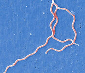
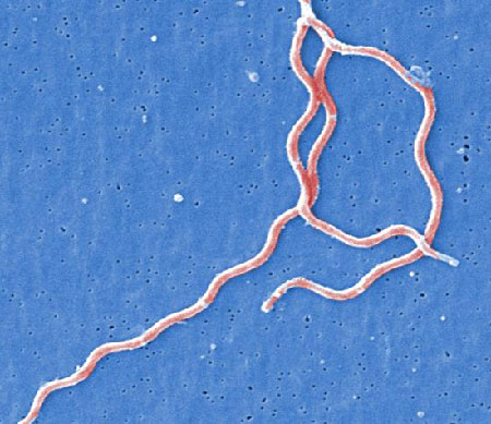
Under a high magnification, this digitally-colorized scanning electron micrograph depicts three gram-negative, anaerobic, Borrelia burgdorferi bacteria, which had been derived from a pure culture
CDC Image Library
See this image in context in the following section/s:
Lyme disease
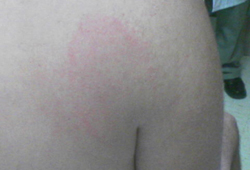
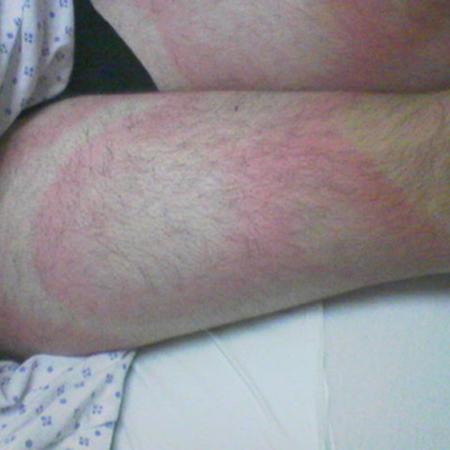
Erythema migrans
From the personal collection of Dr Cristian Speil; used with permission
See this image in context in the following section/s:
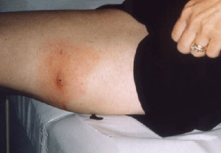
Lyme disease
Erythema migrans
From the personal collection of Dr Cristian Speil; used with permission
See this image in context in the following section/s:

Lyme disease
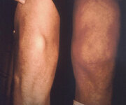
This Lyme disease patient presented with the signs and symptoms indicative of arthritic changes to his right knee due to a Borrelia burgdorferi bacterial infection
CDC Image Library
See this image in context in the following section/s:

Lyme disease
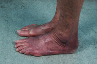
Acrodermatitis chronica atrophicans
Nguyen AL et al. Case Reports 2016; 2016: bcr2016216033; used with permission
See this image in context in the following section/s:
Lyme disease
Lateral aspect of the left thigh of a patient who’d presented with what was diagnosed as Lyme disease showing the characteristic red, expanding rash (erythema migrans)
CDC Image Library
See this image in context in the following section/s:

Lyme disease
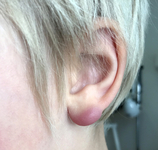
Borrelial lymphocytoma
Gzzz, CC BY-SA 4.0 via Wikimedia Commons; used with permission
See this image in context in the following section/s:

Lyme disease
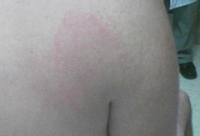
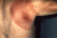
Posterior right shoulder region of a patient with Lyme disease showing erythema migrans
CDC Image Library
See this image in context in the following section/s:
Lyme disease
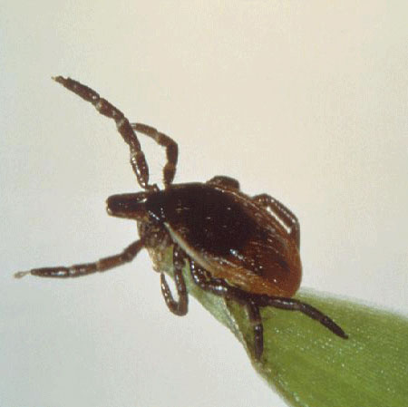
Deer tick (or blacklegged tick), Ixodes scapularis, as it was questing on a blade of grass
CDC Image Library
See this image in context in the following section/s:
Use of this content is subject to our disclaimer