Images and videos
Images


Glomerulonephritis
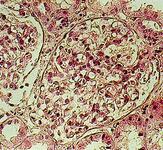
Minimal change nephropathy: the glomerulus has a normal appearance
From: Mason PD, Musey CD. BMJ. 1994 Dec 10;309(6968):1557-63
See this image in context in the following section/s:
Glomerulonephritis
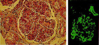
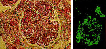
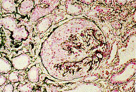
Mesangiocapillary glomerulonephritis showing (left) thickened capillary loops with diffuse cellular proliferation, giving characteristic "lobular" appearance (light microscopy; stains: hematoxylin and eosin) and (right) coarse patchy granular immunofluorescent staining of IgM along capillary loops
From: Mason PD, Musey CD. BMJ. 1994 Dec 10;309(6968):1557-63
See this image in context in the following section/s:
Glomerulonephritis
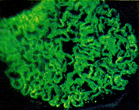
Membranous nephropathy showing fine granular immunofluorescent staining of IgG along basement membrane
Mason PD, Musey CD. BMJ. 1994 Dec 10;309(6968):1557-63
See this image in context in the following section/s:
Glomerulonephritis
Crescentic glomerulonephritis with cellular crescent occupying large portion Bowman's capsule and compressing glomerular tuft
From: Mason PD, Musey CD. BMJ. 1994 Dec 10;309(6968):1557-63
See this image in context in the following section/s:
Glomerulonephritis
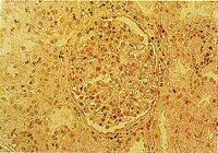
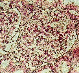
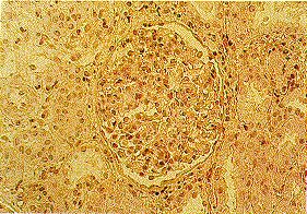
Diffuse proliferative glomerulonephritis as seen in poststreptococcal glomerulonephritis
From: Mason PD, Musey CD. BMJ. 1994 Dec 10;309(6968):1557-63
See this image in context in the following section/s:

Glomerulonephritis
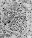
Light microscopy of kidney biopsy showing typical lesions of focal segmental glomerulosclerosis (arrows)
Adapted from Nagi AH, Alexander F, Lannigan R. J Clin Pathol. 1971 Dec;24(9):846-50
See this image in context in the following section/s:
Glomerulonephritis
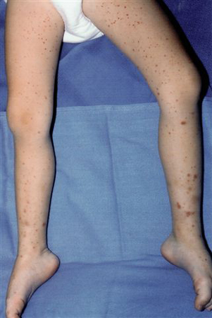
Palpable purpura on the lower extremities of a child with IgA vasculitis (Henoch-Schonlein purpura)
From the collection of Dr Paul F. Roberts, Mayo Clinic
See this image in context in the following section/s:
Glomerulonephritis
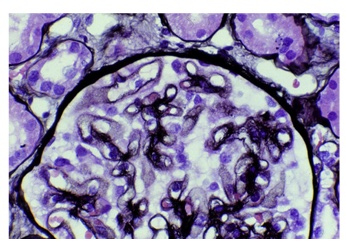
Membranous nephropathy with thickened capillary walls with the appearance of numerous pinpoint “holes” in tangential sections, indicating deposits that did not stain (light microscopy; Jones silver stain)
Fogo AB, Lusco MA, Najafian B, et al. Am J Kidney Dis. 2015;66(3):e15-7
See this image in context in the following section/s:

Glomerulonephritis
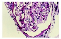
Membranous nephropathy shows slightly prominent capillary walls that appear more rigid than normal; however, deposits cannot be directly visualized (light microscopy; periodic acid Schiff stain)
Fogo AB, Lusco MA, Najafian B, et al. Am J Kidney Dis. 2015;66(3):e15-7
See this image in context in the following section/s:
Use of this content is subject to our disclaimer