Images and videos
Images

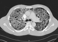

Pneumocystis jirovecii pneumonia
Computed tomography scan of the thorax showing bilateral pulmonary interstitial infiltrates and pneumatoceles (cysts), which are typical of Pneumocystis pneumonia (PCP)
From the collection of Matthew Gingo, UPMC
See this image in context in the following section/s:
Pneumocystis jirovecii pneumonia
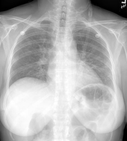
Posteroanterior chest x-ray showing mild, reticular, bilateral pulmonary interstitial infiltrates
From the collection of Matthew Gingo, UPMC
See this image in context in the following section/s:
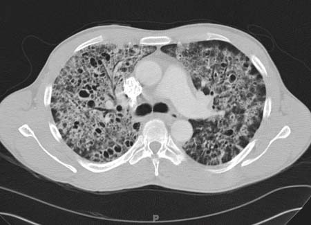
Pneumocystis jirovecii pneumonia
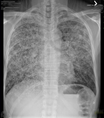
Posteroanterior chest x-ray showing severe, bilateral pulmonary interstitial infiltrates with pneumatoceles
From the collection of Matthew Gingo, UPMC
See this image in context in the following section/s:
Pneumocystis jirovecii pneumonia
Algorithm for the diagnosis of Pneumocystis pneumonia; bronchoalveolar lavage (BAL)
Matthew Gingo, adapted from Singh, HIV Clinical Manual, 2003
See this image in context in the following section/s:
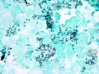
Pneumocystis jirovecii pneumonia
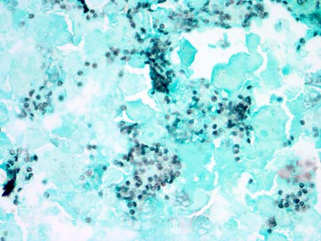
Photomicrograph of bronchoalveolar lavage (BAL) fluid showing Pneumocystis cysts, stained black with Grocott-Gomori methenamine-silver stain (methyl green counterstain)
From the collection of Matthew Gingo, UPMC
See this image in context in the following section/s:
Use of this content is subject to our disclaimer