Aetiology
Pneumocystis pneumonia (PCP) is caused by the fungal organism Pneumocystis jirovecii (formerly Pneumocystis carinii). The mode of transmission of Pneumocystis is not well understood, but airborne transmission is likely on the basis of animal and human studies.[32]
Pneumonia in humans caused by Pneumocystis was first recognised in the 1940s and 1950s. The organism was thought to be a protozoan until 1988 when RNA studies showed greater homology with fungi.[33] In 2001, Pneumocystis was re-classified as a fungus and the organism re-named Pneumocystis jirovecii.[34][35]Pneumocystis pneumonia is still referred to most commonly as PCP.
Pathophysiology
Infection in the lung starts with the multiplication of the organism in the alveoli. As infection progresses, alveoli fill with exudates, there is type 2 pneumocyte hyperplasia, and mononuclear cells infiltrate the lung. Desquamation of the alveolar lining cells creates increased permeability of the alveolar capillary membrane and non-cardiogenic pulmonary oedema.
In patients with AIDS, there are larger numbers of Pneumocystis organisms in the lungs and fewer inflammatory cells than in patients with PCP who are HIV-negative.[36] Fewer inflammatory cells in AIDS patients with PCP seems to be related to better oxygenation and survival than in patients without AIDS.[36]
Further lung destruction can lead to formation of pneumatoceles (also known as cysts, blebs, or cavities) and pneumothoraces. This damage may occur from chronic, low-grade infection, erosion of lung parenchyma from inflammation causing release of proteases and elastases, or alteration in lung connective tissue by HIV infection.[37][Figure caption and citation for the preceding image starts]: Posteroanterior chest x-ray showing severe, bilateral pulmonary interstitial infiltrates with pneumatocelesFrom the collection of Matthew Gingo, UPMC [Citation ends].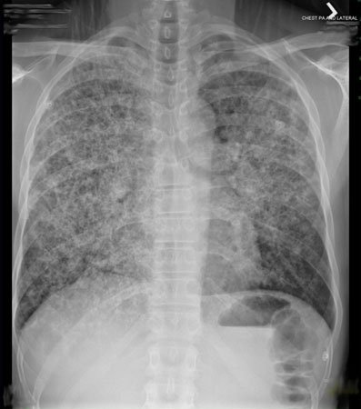 [Figure caption and citation for the preceding image starts]: Computed tomography scan of the thorax showing bilateral pulmonary interstitial infiltrates and pneumatoceles (cysts), which are typical of Pneumocystis pneumonia (PCP)From the collection of Matthew Gingo, UPMC [Citation ends].
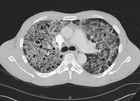
Classification
Biological classification
Kingdom: Fungi
Sub-kingdom: Dikarya
Phylum: Ascomycota
Subphylum: Taphrinomycotina
Class: Pneumocystidomycetes
Order: Pneumocystidales
Family: Pneumocystidaceae
Genus: Pneumocystis
Use of this content is subject to our disclaimer