Images and videos
Images


Multiple sclerosis
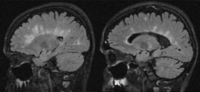
Sagittal fluid-attenuated inversion recovery (FLAIR) images with typical MS lesions involve the corpus callosum either as discrete lesions or as finger-like projections perpendicular to the corpus callosum. Note also the enlargement of the ventricles and diffuse atrophy of more advanced MS
From the collection of Dr Lael A. Stone
See this image in context in the following section/s:
Multiple sclerosis
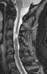
Cervical spine magnetic resonance imaging scan illustrating neuromyelitis optica spectrum disorder. Extensive multiple levels of cervical spinal cord involvement with edema and blood-brain barrier breakdown as illustrated by the contrast-enhanced T1-weighted image (left). The T2-weighted image (right) indicates the extent of signal abnormality that may manifest clinically as quadriparesis with severe spasticity and pain
From the collection of Dr Lael A. Stone
See this image in context in the following section/s:

Multiple sclerosis
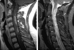
Magnetic resonance imaging scan of the cervical spine at high (≥1 Tesla) magnetic field strength illustrating a lesion that may cause myelopathic symptoms of bowel and bladder dysfunction, as well as spastic paraparesis
From the collection of Dr Lael A. Stone
See this image in context in the following section/s:
Multiple sclerosis
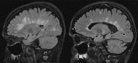
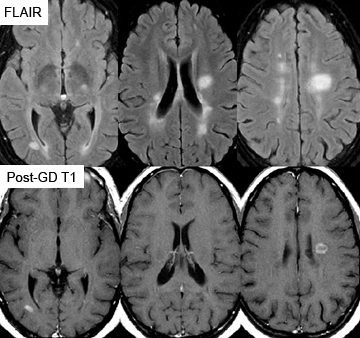
Representative axial magnetic resonance images using fluid-attenuated inversion recovery (FLAIR) showing typical lesions seen in MS in the periventricular regions. Comparable slices using the contrast agent gadolinium illustrate blood-brain barrier breakdown/active inflammation in 2 of the lesions. The FLAIR lesions that do not enhance are likely to be older, with a combination of gliosis and low-level chronic inflammation and degeneration
From the collection of Dr Lael A. Stone
See this image in context in the following section/s:
Videos
 Diagnostic lumbar puncture in adults: animated demonstration
How to perform a diagnostic lumbar puncture in adults. Includes a discussion of patient positioning, choice of needle, and measurement of opening and closing pressure.
 Venepuncture and phlebotomy: animated demonstration
How to take a venous blood sample from the antecubital fossa using a vacuum needle.
 Peripheral intravascular catheter: animated demonstration
How to insert a peripheral intravascular catheter into the dorsum of the hand.
Use of this content is subject to our disclaimer