Images and videos
Images


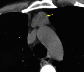

Thymic tumor
CT scan of the chest showing Masaoka-Koga stage III thymoma with abutment of the anterior chest wall and invasion of the medial aspect of the left lung
From the collection of Cameron Wright, MD; used with permission
See this image in context in the following section/s:
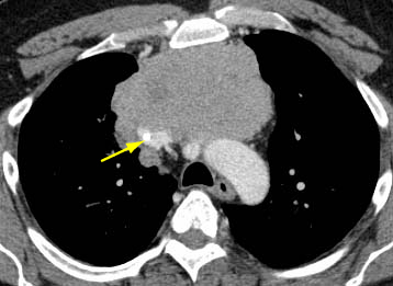
Thymic tumor
CT scan of the chest showing thymoma with encasement and invasion of the superior vena cava
From the collection of Cameron Wright, MD; used with permission
See this image in context in the following section/s:

Thymic tumor
CT scan of the chest showing thymoma with encasement and invasion of the left innominate vein
From the collection of Cameron Wright, MD; used with permission
See this image in context in the following section/s:

Thymic tumor
CT scan of the chest showing a prominent thymic gland with bilobed appearance, consistent with thymic hyperplasia
From the collection of Cameron Wright, MD; used with permission
See this image in context in the following section/s:
Thymic tumor
CT scan of the chest showing a typical Masaoka-Koga stage I thymoma
From the collection of Cameron Wright, MD; used with permission
See this image in context in the following section/s:

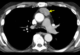
Thymic tumor
CT scan of the chest showing Masaoka-Koga stage IVA thymoma with pleural effusion (red arrow) and extensive pleural metastases (yellow arrow) along posterior chest wall
From the collection of Cameron Wright, MD; used with permission
See this image in context in the following section/s:
Use of this content is subject to our disclaimer