Images and videos
Images


Orbital fractures
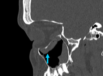
Right orbital blowout fracture on CT-scan; sagittal reformat
From the personal collection of Dr Alistair Cobb
See this image in context in the following section/s:

Orbital fractures
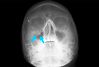
Right orbital blowout fracture; teardrop sign on occipitomental 15° x-ray
From the personal collection of Dr Alistair Cobb
See this image in context in the following section/s:

Orbital fractures
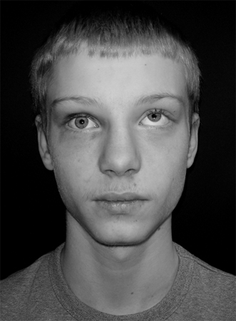
Pediatric blowout fracture: patient looking in central gaze. Mild right circumorbital ecchymosis is noted
From Cobb A, et al. Emerg Med J. 2009;26:351-353; used with permission
See this image in context in the following section/s:
Orbital fractures
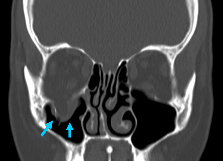
Right orbital blowout fracture on CT-scan; coronal reformat
From the personal collection of Dr Alistair Cobb
See this image in context in the following section/s:
Orbital fractures
Orbital bone anatomy
From Whitaker RH, Borley NR. Instant Anatomy. 3rd ed. Oxford, UK: Blackwell; 2005; used with permission
See this image in context in the following section/s:

Orbital fractures
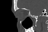
No evidence of fracture on the left orbit as seen on CT-scan; sagittal reconstruction
From the personal collection of Dr Alistair Cobb
See this image in context in the following section/s:
Orbital fractures
Pediatric blowout fracture: patient looking in upward gaze. Right globe limitation by inferior soft tissue entrapment is noted
From Cobb A, et al. Emerg Med J. 2009;26:351-353; used with permission
See this image in context in the following section/s:
Use of this content is subject to our disclaimer