Tests
1st tests to order
visual acuity tests (by specialist)
Test
Verbal children are best tested with multiple targets, or with crowding bars around individual targets.[29] Crowding bars consist of stripes that surround an individual target and thereby simulate the presentation of multiple targets.
Result
reduced, and does not normalize with correction of refractive error alone
stereopsis (perception of depth or 3-dimensionality) and binocular vision testing
Test
The Titmus, Frisby, and Lang stereotests are commonly used to measure stereopsis.
During the Titmus test, patients wear polarized glasses and are asked to identify 3-dimensional images that appear to pop-up toward them. Both the Frisby and Lang stereotests can be performed in free-space without the need for dissociative glasses.
Degree of stereopsis is reported as the number of arc seconds, with lower numbers (40 arc seconds) indicating better stereopsis than higher numbers (3000 arc seconds).
Result
frequently reduced
assessment of fixation, ocular alignment and ocular motility
Test
Patients with a clear visual axis to the fovea and with a normally positioned fovea fixate with the center of the eye, whereas patients with amblyopia, an opacity within the visual axis, or displacement or disease involving the fovea may fixate eccentrically, as if looking at an object from the side.
Marked eccentric fixation is detected by observing the noncentral position of the corneal reflection in the amblyopic eye while the amblyopic eye fixates on a light.
A strabismic patient who can freely alternate fixation between the eyes does not have amblyopia. When either eye is deviated, the fixating other eye should be able to maintain fixation through a blink.
The ocular motility exam may help discover certain patterns of eye movement abnormalities that occur in some children with strabismic amblyopia, although many patients with strabismic amblyopia have full and normal ocular motility.
Result
fixation may be central or eccentric; ocular alignment tests may reveal strabismus; ocular motility may be abnormal
anterior segment exam using a slit lamp
Test
Helps to rule out ocular pathologies that may contribute to decreased vision (e.g., cataracts).
Sometimes, these structural ocular defects are solely responsible for vision loss. However, when they occur asymmetrically or monocularly, coexistant amblyopia may explain a portion of the vision loss.[Figure caption and citation for the preceding image starts]: Portable slit lampFrom the collection of Tina Rutar, MD [Citation ends].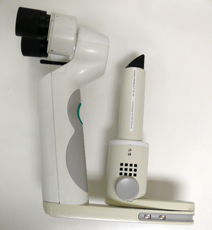 [Figure caption and citation for the preceding image starts]: Slit lampFrom the collection of Tina Rutar, MD [Citation ends].
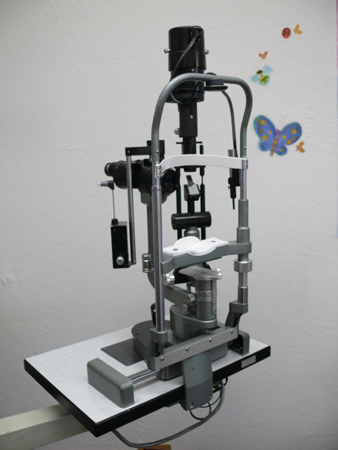 [Figure caption and citation for the preceding image starts]: Author performing slit lamp examFrom the collection of Tina Rutar, MD [Citation ends].
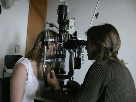
Result
may reveal ocular pathologies contributing to decreased vision (e.g., cataract)
dilated fundoscopy
Test
Typically, cyclopentolate 0.2%/phenylephrine 1% for infants, and cyclopentolate 1% (occasionally with phenylephrine 2.5%) for toddlers and older children.
Helps rule out ocular pathologies that may contribute to decreased vision.
Sometimes, these structural ocular defects are solely responsible for vision loss. However, when they occur asymmetrically or monocularly, coexistant amblyopia may explain a portion of the vision loss.[Figure caption and citation for the preceding image starts]: Indirect ophthalmoscope with 28 diopter lens for performing fundus examFrom the collection of Tina Rutar, MD [Citation ends].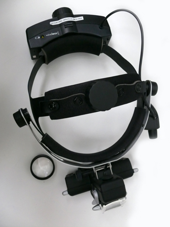 [Figure caption and citation for the preceding image starts]: Author performing indirect ophthalmoscopyFrom the collection of Tina Rutar, MD [Citation ends].
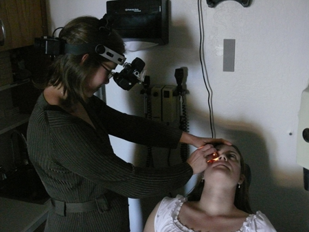
Result
may reveal ocular pathologies contributing to decreased vision (e.g., macular lesions)
cycloplegic retinoscopy
Test
Essential part of complete ophthalmic exam of a child.
Eye drops, typically cyclopentolate 0.2%/phenylephrine 1% for infants, and cyclopentolate 1% for toddlers and children, are used to dilate the pupil and relax the ciliary muscle. Dilated ciliary muscle impairs the strong focusing ability of the child's eye, allowing objective determination of the child's refractive state.
Many causes of amblyopia can only be assessed with a reliable cycloplegic retinoscopy.
At the same time, the quality of the light reflex is assessed to insure nothing in the visual axis interferes with a clear image reaching the retina.[Figure caption and citation for the preceding image starts]: Retinoscope with plus and minus spherical lenses for refractionFrom the collection of Tina Rutar, MD [Citation ends].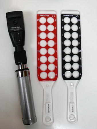
Result
abnormal refractive state, dull retinoscopic reflex, or partially opacified retinoscopic reflex may be present
binocular red reflex test (Brückner test)
Test
Useful screening test for nonophthalmologists. The direct ophthalmoscope light is directed at both pupils from an arm's length away, while the child sits in a darkened room. Patients with an absent or irregular red reflex, or opacity within the reflex, should be referred for ophthalmology assessment promptly, as this could indicate visual axis obstruction from causes such as cataract or intraocular tumor.
Significant hyperopia presents as an inferiorly placed brighter crescent in the red reflex, whereas significant myopia presents as a superiorly placed brighter crescent.[1] Asymmetric crescents could indicate unequal refractive errors between the eyes, which also require further evaluation.
Result
may show media opacities, strabismus, or high refractive error
Emerging tests
visual evoked potentials
Test
Measured using occipital scalp electrodes that detect activity in the visual cortex while visual stimuli are presented to the eyes.
Studies using different types of visual evoked potentials (VEPs): orientation-specific, steady-state motion, multifocal, pattern, and sweep VEPs show that these may be used to predict response to amblyopia therapy.[30][31][32][33][34]
Result
still to be clearly defined, as test is emerging
Use of this content is subject to our disclaimer