History and exam
Key diagnostic factors
uncommon
infant not tracking parent's face
May be due to inadequate vision.
Important feature of history particularly in infants ages 3-6 months.
abnormal red reflex
Abnormalities on exam with a direct ophthalmoscope include asymmetry in color or brightness, absence of reflex, or opacity noted within the reflex.
Although rare, if these abnormalities are present, the child is at risk of form-deprivation amblyopia. It is critical to diagnose in early infancy and to refer for ophthalmology assessment urgently.
Other diagnostic factors
common
asymptomatic
There may be no complaint of any problems with vision. Amblyopia may be detected on routine childhood screening.
subnormal visual acuity for age in one or both eyes
For verbal children (more than about 3 years) Snellen letters are most accurate, but if the child does not know the letters, there are many other options (e.g., Snellen numbers, "H, O, T, V" letters, Lea symbols, Wright figures, and Allen figures).
Indications for referral to an ophthalmologist include visual acuity in either eye worse than 20/50 in a 3-year-old child, visual acuity in either eye worse than 20/40 in a 4- to 5-year-old, or a >2-line difference in visual acuity between the two eyes.[24]
asymmetric corneal light reflex
A light is shone on the eyes from an arm's length away while the patient looks at a small toy held adjacent to the light. If the reflection of the light is symmetrically centered on the cornea in each eye, no manifest strabismus is present. If it is decentered in one eye, manifest strabismus is likely. [Figure caption and citation for the preceding image starts]: Esotropia: left eye fixating (note decentered light reflection on right cornea)From the collection of Daniel J. Salchow, MD [Citation ends].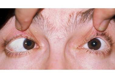
unequal behavioral response to alternate eye occlusion
With normal vision the response should be equal on both sides (i.e., the infant should object or not object equally).
abnormal cover/uncover testing
Test performed if the child is cooperative to testing.
While the child is fixating (staring) at an object, the examiner should cover one eye and look for a refixation movement of the uncovered eye.
Then, the cover should be removed and placed over the other eye to again look for a refixation movement of the uncovered eye. A refixation movement in either eye indicates strabismus.
Children with strabismus require referral to an ophthalmologist for further assessment.
If the child constantly fixates with one eye and the other eye remains deviated, vision is decreased in the deviated eye, which is often due to strabismic amblyopia.
Strabismic children who alternate fixation between the two eyes and can maintain fixation through a blink with either eye probably do not have amblyopia.
uncommon
blurred vision
May indicate high refractive error or high astigmatism, which may result in amblyopia.
eye strain
May be a complaint but is not a common presentation of amblyopia.[4]
congenital nystagmus
Nystagmus occurring before age 6 months can be due to sensory and motor abnormalities of the visual system.
Bilateral form-deprivation amblyopia causes a bilateral sensory nystagmus. It portends an unfavorable visual outcome, suggesting that amblyopia treatment has already been delayed past the optimal time period.
abnormal pupil exam
May indicate ocular pathologies that may be contributing to decreased vision (e.g., abnormal pupillary response may be due to optic nerve hypoplasia or retinal dystrophy). May be the sole cause of decreased vision or may be associated with amblyopia, especially if unilateral.
Children or infants with abnormalities in pupillary shape, pupils of unequal size, or poor and unequal reaction to light should be referred to an ophthalmologist.
abnormal external eye exam
Children or infants who have structural abnormalities of the eye, eyelids, or orbit detected through external inspection should be referred to an ophthalmologist for further investigation. A penlight is sufficient for external inspection of the eye.
Severe ptosis is associated with developing form-deprivation amblyopia.
Risk factors
strong
age <9 years
The developing brain of a young child is most susceptible to abnormal visual stimulation. The older the child, the more resistant that child is to the effects of optical defocus, suppression, and visual deprivation.
Although amblyopia develops during childhood, the effects of untreated amblyopia last a lifetime. Some adults can regain vision in the amblyopic eye after loss of vision in the previously normal eye, but this type of plasticity in the adult visual system is uncommon.[12][13]
strabismus (misalignment of the eyes)
Thought to result in amblyopia due to the competitive or inhibitory interaction between cortical inputs from the two eyes. Cortical vision centers from the fixating eye dominate, and cortical vision centers from the nonfixating eye are chronically suppressed.[1]
opacity in the cornea, anterior chamber, lens, vitreous, or retinal surface
Such as congenital cataract, corneal opacity, nonclearing vitreous hemorrhage, and macular hemorrhage.
Blurring of the retinal image may lead to form-deprivation amblyopia.
Unilateral occlusion tends to be worse than bilateral occlusion of similar magnitude because interocular competition adds to the direct developmental impact of severe image degradation.[1]
Central lenticular opacities >3 mm in size are amblyogenic.[14][15]
severe ptosis or prolonged occlusion of one or both eyes
May result in form-deprivation amblyopia.
weak
prematurity
Premature infants, compared with full-term infants, have an increased risk of long-term decreased visual acuity that is independent of factors such as retinopathy of prematurity and neurologic disorders.[16]
Compared with full-term infants, premature infants have a higher likelihood of refractive error and of strabismus, both of which can cause amblyopia.[17] Premature children treated with laser for retinopathy of prematurity (ROP) have a high incidence of myopia or high myopia.[18][19][20] The Early Treatment of Retinopathy of Prematurity (ETROP) study found that, at age 3 years, the prevalence of myopia in infants with severe ROP was 65% to 71% and the prevalence of high myopia (≤-5.00 diopter) was 51%.[19] The rates of hyperopia and astigmatism in children born prematurely are also higher compared to children born full-term.[21]
Some conditions that cause form-deprivation amblyopia (e.g., congenital cataract) occur more frequently in infants born prematurely. These infants may have genetic syndromes or may have been exposed to intrauterine infections or toxins that have contributed to an early delivery.
family history of amblyopia or strabismus
Although amblyopia and strabismus are not inherited as Mendelian traits, both have a genetic predisposition. Siblings of children with accommodative esotropia (inward turning of the eye on accommodation; a type of strabismus that commonly leads to amblyopia) showed a high prevalence of amblyogenic risk factors, including strabismus, moderate to high hyperopia, moderate to high astigmatism, and anisometropia.[22]
Risk of accommodative esotropia was nearly 50% among children who had moderate to high hyperopia and whose parents had a history of strabismus.[23] Accommodative esotropia is a common cause of amblyopia.
hyperopic anisometropia (farsightedness with unequal refractive error between the 2 eyes) of >+1.50 diopter
The child focuses with the less hyperopic eye both at distance and at near, resulting in chronic defocus of the more hyperopic eye.
The difference in hyperopia between the two eyes that is necessary to induce amblyopia is not known, but generally >+1.50 diopter difference is required.[1]
There is a correlation between amount of anisometropia and depth of amblyopia.
myopic anisometropia
Can cause amblyopia, but greater differences between the power of the two eyes are needed than with hyperopic anisometropia. This association is less common than that of hyperopic anisometropia with amblyopia.
People with myopia have a focal point near where images are clear. If the two focal points are different for the right and left eyes, the child may prefer one eye for one viewing distance and the other eye for another viewing distance. There is not a chronic imbalance leading to favoritism for one eye at all viewing distances.
astigmatism (cylindrical, rather than spherical, defocus of the eye) >2.00 diopter
Most normal children have some astigmatism; astigmatism of up to approximately 1.50 diopter is associated with normal vision and visual development. However, high astigmatism can cause meridional amblyopia.
Astigmatism occurring in the vertical and horizontal meridian induces less image blur than astigmatism at oblique meridians. In children ages 2 years and older, vertical or horizontal astigmatism of approximately 2.00 diopter may be amblyogenic, and oblique astigmatism of approximately 1.50 diopter may be amblyogenic.[1]
hyperopia >+4.50 diopter
Although mild to moderate hyperopia in the absence of strabismus is not amblyogenic, high hyperopia can cause amblyopia.[1]
Although ciliary muscles in childhood are powerful and can focus the lens of the eye many diopters, children may not completely bring images into focus if exceptional focusing effort is required. A hyperopic child may be perfectly content seeing the world slightly blurred, and the chronic defocus can lead to amblyopia.
myopia >-3.00 diopter
High myopia is better tolerated than high hyperopia because those with high myopia still have a focal point near where they can see clearly with no focusing effort. Infants and toddlers are initially more interested in the near world than the distant world, so their visual development is usually unaffected by myopia. However, high myopia has a very near focal point, which may be impractical (e.g., at -10.0 diopter myopia the focal point is at 10 cm, and even an infant or a toddler may not get enough visual stimulation focusing on objects so close to the surface of the eye).
Practice guidelines include myopia of ≥-3.00 diopter as potentially amblyogenic.[1]
developmental delay
Amblyopia may occur more commonly among children with developmental delay.[1] These children may be unable to cooperate with visual acuity testing in a pediatrician’s office or during school or community-based vision screenings. If they cannot be screened, they should be referred to a pediatric ophthalmologist or optometrist for a complete evaluation.
Use of this content is subject to our disclaimer