Tests
1st tests to order
culture of pus or affected tissue
Test
Actinomycosis is usually diagnosed by culturing the pathogen from affected tissue under strict anaerobic or at least microaerophilic conditions. Culture requires a minimum of 14 days.[1] Clinicians should specifically request actinomycosis culture on the laboratory request form.[7] False-negative results are not uncommon.
Patients have often already been treated with antibiotics before the differential diagnosis of actinomycosis is considered. In such cases culturing the agent is difficult or impossible. Therefore, although culture of the organism from affected tissue is the preferred diagnostic test, in many cases the diagnosis can made only by histology or immunohistology.
Result
growth of actinomycetes
histology of affected tissue
Test
Sections of biopsies reveal acute or chronic inflammation and granulation. [Figure caption and citation for the preceding image starts]: Periodic acid-Schiff stain of a mass of actinomycetes in a lymph nodeFrom the collection of Professor Dr Christoph Loddenkemper, Department of Pathology, Charité - University Medicine Berlin, CBF, Berlin, Germany [Citation ends].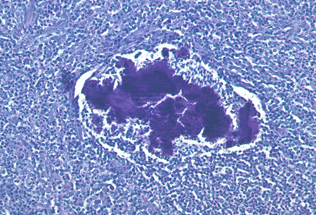 Neutrophils, foamy macrophages, plasma cells, and lymphocytes surrounding dense fibrotic tissue are usually seen.
Actinomyces form characteristic sulfur granules, which are composed of a protein-polysaccharide complex and mineralized by host calcium and phosphate.[35] The name reflects the yellow color of the granule in pus. Sulfur granules may be seen in only a few sections and in low density. Sometimes more than 1 biopsy is needed to confirm the diagnosis by this method.
The granules are often discrete, about 100 to 1000 micrometers in diameter, and are often seen directly without magnification or under the microscope with low magnification.
Result
acute or chronic inflammation and granulation tissue; sulfur granules
immunohistology
Test
Specific staining using fluorescent-conjugated monoclonal antibodies has improved diagnostic procedures.[1]
Result
detection of actinomycetes within affected tissue
CBC
Test
Anemia may result from vaginal blood loss in pelvic actinomycosis. Elevated WBC counts are a common, nonspecific feature in all forms of actinomycosis.
Result
anemia or leukocytosis
CT or MRI of abdomen
Test
May show abdominal and other masses caused by actinomycosis, although these are unlikely to be diagnostic.
Result
visualization of actinomycotic mass
Emerging tests
PCR of affected tissue
Test
No standardized protocols exist. Used mainly in research studies. Has not become a routine diagnostic procedure.
Result
detection of actinomycetes' DNA/RNA
Use of this content is subject to our disclaimer