Although there are several classic symptoms and signs typical of molar pregnancy, such as vaginal bleeding, hyperemesis, or hyperthyroidism, most women with molar pregnancy in modern clinical practice are diagnosed incidentally at histologic exam of the products of miscarriage or on findings from early maternal ultrasound.[30]Lok C, Frijstein M, van Trommel N. Clinical presentation and diagnosis of gestational trophoblastic disease. Best Pract Res Clin Obstet Gynaecol. 2021 Jul;74:42-52.
https://www.sciencedirect.com/science/article/abs/pii/S1521693420301760?via%3Dihub
http://www.ncbi.nlm.nih.gov/pubmed/33422446?tool=bestpractice.com
[31]American College of Obstetricians and Gynecologists. ACOG practice bulletin no. 175: ultrasound in pregnancy. Dec 2016 [internet publication].
https://www.acog.org/clinical/clinical-guidance/practice-bulletin/articles/2016/12/ultrasound-in-pregnancy
History
Women with molar pregnancy typically present in the first trimester of pregnancy with a history of a missed menstrual period, and are found to have positive urine test for pregnancy and elevated serum human chorionic gonadotropin (hCG) levels on laboratory evaluation. Women are commonly at the extremes of reproductive life (younger than 20 years of age or over 35 years of age), and may relate a history of prior MP.[8]Sebire NJ, Fisher RA, Foskett M, et al. Risk of recurrent hydatidiform mole and subsequent pregnancy outcome following complete or partial hydatidiform molar pregnancy. BJOG. 2003 Jan;110(1):22-6.
http://www.ncbi.nlm.nih.gov/pubmed/12504931?tool=bestpractice.com
[9]Vargas R, Barroilhet LM, Esselen K, et al. Subsequent pregnancy outcomes after complete and partial molar pregnancy, recurrent molar pregnancy, and gestational trophoblastic neoplasia: an update from the New England Trophoblastic Disease Center. J Reprod Med. 2014 May-Jun;59(5-6):188-94.
http://www.ncbi.nlm.nih.gov/pubmed/24937955?tool=bestpractice.com
[10]Altman AD, Bentley B, Murray S, et al. Maternal age-related rates of gestational trophoblastic disease. Obstet Gynecol. 2008 Aug;112(2 Pt 1):244-50.
http://www.ncbi.nlm.nih.gov/pubmed/18669718?tool=bestpractice.com
[11]Savage PM, Sita-Lumsden A, Dickson S, et al. The relationship of maternal age to molar pregnancy incidence, risks for chemotherapy and subsequent pregnancy outcome. J Obstet Gynaecol. 2013 May;33(4):406-11.
https://www.tandfonline.com/doi/abs/10.3109/01443615.2013.771159
http://www.ncbi.nlm.nih.gov/pubmed/23654327?tool=bestpractice.com
[12]Gockley AA, Melamed A, Joseph NT, et al. The effect of adolescence and advanced maternal age on the incidence of complete and partial molar pregnancy. Gynecol Oncol. 2016 Mar;140(3):470-3.
http://www.ncbi.nlm.nih.gov/pubmed/26777992?tool=bestpractice.com
[13]Di Cintio E, Parazzini F, Rosa C, et al. The epidemiology of gestational trophoblastic disease. Gen Diagn Pathol. 1997 Nov;143(2-3):103-8.
http://www.ncbi.nlm.nih.gov/pubmed/9443567?tool=bestpractice.com
[14]Braga A, Growdon WB, Bernstein M, et al. Molar pregnancy in adolescents. J Reprod Med. 2012 May-Jun;57(5-6):225-30.
http://www.ncbi.nlm.nih.gov/pubmed/22696817?tool=bestpractice.com
[15]Soares RR, Maestá I, Colón J, et al. Complete molar pregnancy in adolescents from North and South America: Clinical presentation and risk of gestational trophoblastic neoplasia. Gynecol Oncol. 2016 Sep;142(3):496-500.
http://www.ncbi.nlm.nih.gov/pubmed/27423380?tool=bestpractice.com
[32]Tranoulis A, Georgiou D, Sayasneh A, et al. Gestational trophoblastic neoplasia: a meta-analysis evaluating reproductive and obstetrical outcomes after administration of chemotherapy. Int J Gynecol Cancer. 2019 Jul;29(6):1021-31.
http://www.ncbi.nlm.nih.gov/pubmed/31253638?tool=bestpractice.com
The most common presenting symptom is vaginal bleeding.[33]Beguin Y, Huebers HA, Josephson B, et al. Transferrin receptors in rat plasma. Proc Natl Acad Sci U S A. 1988 Jan;85(2):637-40.
https://www.pnas.org/doi/10.1073/pnas.85.2.637?url_ver=Z39.88-2003&rfr_id=ori%3Arid%3Acrossref.org&rfr_dat=cr_pub++0pubmed
http://www.ncbi.nlm.nih.gov/pubmed/3422446?tool=bestpractice.com
[34]Tidy, J, Seckl, M, Hancock, BW, on behalf of the Royal College of Obstetricians and Gynaecologists. Management of gestational trophoblastic disease. BJOG 2021;128: e1-e27.
https://obgyn.onlinelibrary.wiley.com/doi/full/10.1111/1471-0528.16266
This may vary in its degree from light spotting to heavy bleeding, and may even include passage of hydropic villi. Heavy or persistent bleeding can lead to anemia, with symptoms of dizziness and fatigue.
Women may report exacerbated symptoms of pregnancy (as a result of abnormally high serum hCG levels) that include severe nausea and emesis (hyperemesis gravidarum), palpitations, insomnia and diarrhea (from thyrotoxicity), and headache and photophobia (from early-onset preeclampsia).[24]Braga A, Moraes V, Maestá I, et al. Changing trends in the clinical presentation and management of complete hydatidiform mole among Brazilian women. Int J Gynecol Cancer. 2016 Jun;26(5):984-90.
http://www.ncbi.nlm.nih.gov/pubmed/26905335?tool=bestpractice.com
[25]Sun SY, Melamed A, Goldstein DP, et al. Changing presentation of complete hydatidiform mole at the New England Trophoblastic Disease Center over the past three decades: does early diagnosis alter risk for gestational trophoblastic neoplasia? Gynecol Oncol. 2015 Jul;138(1):46-9.
http://www.ncbi.nlm.nih.gov/pubmed/25969351?tool=bestpractice.com
[35]Soto-Wright V, Bernstein M, Goldstein DP, et al. The changing clinical presentation of complete molar pregnancy. Obstet Gynecol. 1995 Nov;86(5):775-9.
http://www.ncbi.nlm.nih.gov/pubmed/7566847?tool=bestpractice.com
High-output cardiac failure from hyperthyroidism, severe preeclampsia and, less commonly, anemia may lead to dyspnea and respiratory distress. Dyspnea may also indicate trophoblastic emboli or pulmonary metastases. Because it is accompanied by significantly higher levels of serum hCG, complete hydatidiform mole is more likely to be associated with these symptoms than partial molar pregnancy.[23]Berkowitz RS, Goldstein DP. Chorionic tumors. N Engl J Med. 1996 Dec 5;335(23):1740-8.
http://www.ncbi.nlm.nih.gov/pubmed/8929267?tool=bestpractice.com
Women with molar pregnancies may also experience pelvic pain secondary to ovarian theca lutein cysts.
Hydatidiform moles with severe medical complications occur less frequently because of early diagnosis from the widespread use of ultrasound in the first trimester of pregnancy. About 50% of women with molar pregnancies are diagnosed while still asymptomatic. However, earlier diagnosis has not been associated with a lower occurrence of postmolar gestational trophoblastic neoplasia (GTN), and prognosis is still a clinical challenge.[24]Braga A, Moraes V, Maestá I, et al. Changing trends in the clinical presentation and management of complete hydatidiform mole among Brazilian women. Int J Gynecol Cancer. 2016 Jun;26(5):984-90.
http://www.ncbi.nlm.nih.gov/pubmed/26905335?tool=bestpractice.com
[25]Sun SY, Melamed A, Goldstein DP, et al. Changing presentation of complete hydatidiform mole at the New England Trophoblastic Disease Center over the past three decades: does early diagnosis alter risk for gestational trophoblastic neoplasia? Gynecol Oncol. 2015 Jul;138(1):46-9.
http://www.ncbi.nlm.nih.gov/pubmed/25969351?tool=bestpractice.com
Physical exam
The uterine size is greater than expected for gestational age in approximately 25% of complete molar pregnancies.[36]Sun SY, Melamed A, Joseph NT, et al. Clinical presentation of complete hydatidiform mole and partial hydatidiform mole at a regional trophoblastic disease center in the United States over the past 2 decades. Int J Gynecol Cancer. 2016 Feb;26(2):367-70.
http://www.ncbi.nlm.nih.gov/pubmed/26588240?tool=bestpractice.com
[37]Lurain JR. Hydatidiform mole: recognition and management. Contemporary OB/GYN. 2019;64(3).
https://www.contemporaryobgyn.net/view/hydatidiform-mole-recognition-and-management
There may be active bleeding from the cervical os, and there may be spontaneous evacuation of hydropic vesicles from the cervix.[4]Ngan HYS, Seckl MJ, Berkowitz RS, et al. Diagnosis and management of gestational trophoblastic disease: 2021 update. Int J Gynaecol Obstet. 2021 Oct;155 Suppl 1(suppl 1):86-93.
https://obgyn.onlinelibrary.wiley.com/doi/10.1002/ijgo.13877
http://www.ncbi.nlm.nih.gov/pubmed/34669197?tool=bestpractice.com
Other signs include pallor, tachycardia, tremor, hypertension, and respiratory distress. Because of the molecular homology between subunits of thyroid-stimulating hormone and hCG, serum hCG may stimulate the production of thyroid hormone with the clinical symptoms and signs of thyrotoxicosis.[26]Ramos MM, Maesta I, de Araújo Costa RA, et al. Clinical characteristics and thyroid function in complete hydatidiform mole complicated by hyperthyroidism. Gynecol Oncol. 2022 Apr;165(1):137-42.
http://www.ncbi.nlm.nih.gov/pubmed/35153074?tool=bestpractice.com
[38]Yamazaki K, Sato K, Shizume K, et al. Potent thyrotropic activity of human chorionic gonadotropin variants in terms of 125I incorporation and de novo synthesized thyroid hormone release in human thyroid follicles. J Clin Endocrinol Metab. 1995 Feb;80(2):473-9.
http://www.ncbi.nlm.nih.gov/pubmed/7852507?tool=bestpractice.com
[39]Bhatia N, Mitharwal SM. Hydatidiform mole with uncontrolled hyperthyroidism: an anesthetic challenge. J Anaesthesiol Clin Pharmacol. 2016 Oct-Dec;32(4):537-8.
https://www.ncbi.nlm.nih.gov/pmc/articles/PMC5187630
http://www.ncbi.nlm.nih.gov/pubmed/28096596?tool=bestpractice.com
However, the absence of ophthalmopathy differentiates molar pregnancy from thyrotoxicosis due to Graves disease.[40]Norman RJ, Green-Thompson RW, Jialal I, et al. Hyperthyroidism in gestational trophoblastic neoplasia. Clin Endocrinol (Oxf). 1981 Oct;15(4):395-401.
http://www.ncbi.nlm.nih.gov/pubmed/7318191?tool=bestpractice.com
Laboratory investigations
Most molar pregnancies (MPs) are diagnosed incidentally at pathologic evaluation of a suction evacuation (electrical or manual) specimen for missed abortion or from early maternal ultrasound screening in the first trimester of pregnancy.[31]American College of Obstetricians and Gynecologists. ACOG practice bulletin no. 175: ultrasound in pregnancy. Dec 2016 [internet publication].
https://www.acog.org/clinical/clinical-guidance/practice-bulletin/articles/2016/12/ultrasound-in-pregnancy
Many women who are diagnosed with MP at an early maternal ultrasound screening in the first trimester of pregnancy terminate pregnancy before the development of the classic signs and symptoms.[24]Braga A, Moraes V, Maestá I, et al. Changing trends in the clinical presentation and management of complete hydatidiform mole among Brazilian women. Int J Gynecol Cancer. 2016 Jun;26(5):984-90.
http://www.ncbi.nlm.nih.gov/pubmed/26905335?tool=bestpractice.com
[25]Sun SY, Melamed A, Goldstein DP, et al. Changing presentation of complete hydatidiform mole at the New England Trophoblastic Disease Center over the past three decades: does early diagnosis alter risk for gestational trophoblastic neoplasia? Gynecol Oncol. 2015 Jul;138(1):46-9.
http://www.ncbi.nlm.nih.gov/pubmed/25969351?tool=bestpractice.com
[35]Soto-Wright V, Bernstein M, Goldstein DP, et al. The changing clinical presentation of complete molar pregnancy. Obstet Gynecol. 1995 Nov;86(5):775-9.
http://www.ncbi.nlm.nih.gov/pubmed/7566847?tool=bestpractice.com
For this reason, most authorities consider it mandatory to conduct histologic exam of miscarriage tissue.[4]Ngan HYS, Seckl MJ, Berkowitz RS, et al. Diagnosis and management of gestational trophoblastic disease: 2021 update. Int J Gynaecol Obstet. 2021 Oct;155 Suppl 1(suppl 1):86-93.
https://obgyn.onlinelibrary.wiley.com/doi/10.1002/ijgo.13877
http://www.ncbi.nlm.nih.gov/pubmed/34669197?tool=bestpractice.com
[34]Tidy, J, Seckl, M, Hancock, BW, on behalf of the Royal College of Obstetricians and Gynaecologists. Management of gestational trophoblastic disease. BJOG 2021;128: e1-e27.
https://obgyn.onlinelibrary.wiley.com/doi/full/10.1111/1471-0528.16266
hCG is secreted by syncytiotrophoblasts, which proliferate excessively in molar pregnancy, and so it is a sensitive biomarker for diagnosis of MP, response to treatment and follow-up monitoring for GTN.[22]Soper JT. Gestational trophoblastic disease. Obstet Gynecol. 2006 Jul;108(1):176-87.
http://www.ncbi.nlm.nih.gov/pubmed/16816073?tool=bestpractice.com
Normal pregnancies are associated with a peak serum hCG level of <100,000 mIU/mL.[23]Berkowitz RS, Goldstein DP. Chorionic tumors. N Engl J Med. 1996 Dec 5;335(23):1740-8.
http://www.ncbi.nlm.nih.gov/pubmed/8929267?tool=bestpractice.com
However, hCG levels alone should not be used to infer the presence of a molar pregnancy because greater than expected hCG levels may be seen in multiple gestations. Serum hCG acts as a tumor marker to follow postmolar regression and to identify postmolar gestational trophoblastic neoplasia. In the absence of histopathology, post-treatment measurement of hCG levels should be performed weekly until normalization of hCG levels or diagnosis of GTN. The duration of monitoring varies by country.[3]Horowitz NS, Eskander RN, Adelman MR, et al. Epidemiology, diagnosis, and treatment of gestational trophoblastic disease: A Society of Gynecologic Oncology evidenced-based review and recommendation. Gynecol Oncol. 2021 Dec;163(3):605-13.
https://www.gynecologiconcology-online.net/article/S0090-8258(21)01421-9/fulltext
http://www.ncbi.nlm.nih.gov/pubmed/34686354?tool=bestpractice.com
The International Federation of Gynecology and Obstetrics recommends monitoring hCG every 1-2 weeks post-treatment until levels return to normal, followed by a single confirmatory normal measurement within a month for partial hydatidiform moles and monthly hCG measurements for 6 months for complete hydatidiform moles.[4]Ngan HYS, Seckl MJ, Berkowitz RS, et al. Diagnosis and management of gestational trophoblastic disease: 2021 update. Int J Gynaecol Obstet. 2021 Oct;155 Suppl 1(suppl 1):86-93.
https://obgyn.onlinelibrary.wiley.com/doi/10.1002/ijgo.13877
http://www.ncbi.nlm.nih.gov/pubmed/34669197?tool=bestpractice.com
[41]Coyle C, Short D, Jackson L, et al. What is the optimal duration of human chorionic gonadotrophin surveillance following evacuation of a molar pregnancy? A retrospective analysis on over 20,000 consecutive patients. Gynecol Oncol. 2018 Feb;148(2):254-7.
http://www.ncbi.nlm.nih.gov/pubmed/29229282?tool=bestpractice.com
Anemia may result from heavy or persistent vaginal bleeding and the dilutional effects of increased blood volume. There is a greater risk for serious bleeding at the time of evacuation for MP, with a risk of disseminated intravascular coagulation from blood loss or trophoblastic embolism, although these are the rarest complications of molar pregnancy.
About 15% to 20% of women with complete molar pregnancies and 0.5% to 5% of women with partial molar pregnancies develop GTN and require chemotherapy.[4]Ngan HYS, Seckl MJ, Berkowitz RS, et al. Diagnosis and management of gestational trophoblastic disease: 2021 update. Int J Gynaecol Obstet. 2021 Oct;155 Suppl 1(suppl 1):86-93.
https://obgyn.onlinelibrary.wiley.com/doi/10.1002/ijgo.13877
http://www.ncbi.nlm.nih.gov/pubmed/34669197?tool=bestpractice.com
[42]Lok C, van Trommel N, Massuger L, et al. Practical clinical guidelines of the EOTTD for treatment and referral of gestational trophoblastic disease. Eur J Cancer. 2020 May;130:228-40.
http://www.ncbi.nlm.nih.gov/pubmed/32247260?tool=bestpractice.com
These chemotherapy agents require normal liver and renal function for optimal dosing. Women undergoing evacuation of molar pregnancies are at increased risk of bleeding significant enough to require a blood transfusion. Blood typing ensures that type-appropriate blood can be made available in the event of hemorrhage.
Imaging
Pelvic ultrasound is the mainstay of diagnosis and an essential component in the evaluation of suspected GTN.[31]American College of Obstetricians and Gynecologists. ACOG practice bulletin no. 175: ultrasound in pregnancy. Dec 2016 [internet publication].
https://www.acog.org/clinical/clinical-guidance/practice-bulletin/articles/2016/12/ultrasound-in-pregnancy
[43]American College of Radiology. ACR-ACOG-AIUM-SRU practice parameter for the performance of obstetrical ultrasound. 2018 [internet publication].
http://www.acr.org/~/media/ACR/Documents/PGTS/guidelines/US_Obstetrical.pdf
Typical ultrasound findings for a complete molar pregnancy include a diffuse echogenic pattern described as a snow-storm pattern, which is created by intermingling of hydropic villi and blood clots.[3]Horowitz NS, Eskander RN, Adelman MR, et al. Epidemiology, diagnosis, and treatment of gestational trophoblastic disease: A Society of Gynecologic Oncology evidenced-based review and recommendation. Gynecol Oncol. 2021 Dec;163(3):605-13.
https://www.gynecologiconcology-online.net/article/S0090-8258(21)01421-9/fulltext
http://www.ncbi.nlm.nih.gov/pubmed/34686354?tool=bestpractice.com
[42]Lok C, van Trommel N, Massuger L, et al. Practical clinical guidelines of the EOTTD for treatment and referral of gestational trophoblastic disease. Eur J Cancer. 2020 May;130:228-40.
http://www.ncbi.nlm.nih.gov/pubmed/32247260?tool=bestpractice.com
The presence of a smaller volume of abnormal placenta with partial fetal development, without fetal cardiac activity, is characteristic of a partial molar pregnancy.[3]Horowitz NS, Eskander RN, Adelman MR, et al. Epidemiology, diagnosis, and treatment of gestational trophoblastic disease: A Society of Gynecologic Oncology evidenced-based review and recommendation. Gynecol Oncol. 2021 Dec;163(3):605-13.
https://www.gynecologiconcology-online.net/article/S0090-8258(21)01421-9/fulltext
http://www.ncbi.nlm.nih.gov/pubmed/34686354?tool=bestpractice.com
[42]Lok C, van Trommel N, Massuger L, et al. Practical clinical guidelines of the EOTTD for treatment and referral of gestational trophoblastic disease. Eur J Cancer. 2020 May;130:228-40.
http://www.ncbi.nlm.nih.gov/pubmed/32247260?tool=bestpractice.com
Cystic enlargement of the ovaries may represent theca lutein cysts.[3]Horowitz NS, Eskander RN, Adelman MR, et al. Epidemiology, diagnosis, and treatment of gestational trophoblastic disease: A Society of Gynecologic Oncology evidenced-based review and recommendation. Gynecol Oncol. 2021 Dec;163(3):605-13.
https://www.gynecologiconcology-online.net/article/S0090-8258(21)01421-9/fulltext
http://www.ncbi.nlm.nih.gov/pubmed/34686354?tool=bestpractice.com
[Figure caption and citation for the preceding image starts]: Ultrasound showing multiple cystic areas in the uterine cavity giving a "snowstorm appearance" suggestive of molar pregnancy.Nigam A, Kumari A, Gupta N. Negative urine pregnancy test in a molar pregnancy: is it possible? Case Reports 2014;2014:bcr2014206483. [Citation ends].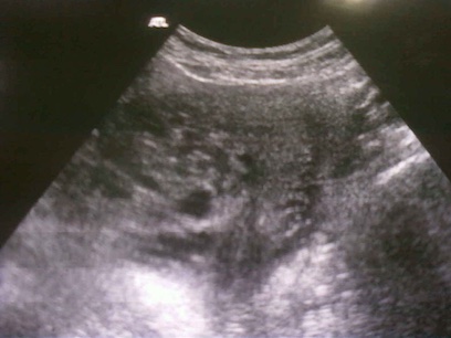
Advanced imaging, such as CT scan or MRI, is not typically required for benign moles, unless the woman has signs or symptoms of pulmonary or brain metastases.[4]Ngan HYS, Seckl MJ, Berkowitz RS, et al. Diagnosis and management of gestational trophoblastic disease: 2021 update. Int J Gynaecol Obstet. 2021 Oct;155 Suppl 1(suppl 1):86-93.
https://obgyn.onlinelibrary.wiley.com/doi/10.1002/ijgo.13877
http://www.ncbi.nlm.nih.gov/pubmed/34669197?tool=bestpractice.com
Women with an established diagnosis of gestational trophoblastic neoplasia (GTN)
The FIGO diagnostic criteria for postmolar GTN without symptoms are based on surveillance of hCG levels and histopathology:[4]Ngan HYS, Seckl MJ, Berkowitz RS, et al. Diagnosis and management of gestational trophoblastic disease: 2021 update. Int J Gynaecol Obstet. 2021 Oct;155 Suppl 1(suppl 1):86-93.
https://obgyn.onlinelibrary.wiley.com/doi/10.1002/ijgo.13877
http://www.ncbi.nlm.nih.gov/pubmed/34669197?tool=bestpractice.com
Four or more measurements of plateaued hCG levels over a 3-week period (on days 1, 7, 14, and 21).
An increase in hCG levels for three or more consecutive weekly measurements over a period of at least 2 weeks (on days 1, 7, and 14).
A histopathologic diagnosis of choriocarcinoma.
Women with an established diagnosis of GTN following molar pregnancy should be evaluated with a pelvic exam and chest x-ray.[3]Horowitz NS, Eskander RN, Adelman MR, et al. Epidemiology, diagnosis, and treatment of gestational trophoblastic disease: A Society of Gynecologic Oncology evidenced-based review and recommendation. Gynecol Oncol. 2021 Dec;163(3):605-13.
https://www.gynecologiconcology-online.net/article/S0090-8258(21)01421-9/fulltext
http://www.ncbi.nlm.nih.gov/pubmed/34686354?tool=bestpractice.com
Pulmonary congestion, edema, alveolar infiltrates, and metastatic nodules may be visible on the chest radiograph. If the chest x-ray is inconclusive, if there are metastases ≥1 cm or the woman has a signs or symptoms of metastatic disease, computed tomography scans of the chest, abdomen, and pelvis should be performed and magnetic resonance imaging of the brain should be obtained to further evaluate and stage metastases.[3]Horowitz NS, Eskander RN, Adelman MR, et al. Epidemiology, diagnosis, and treatment of gestational trophoblastic disease: A Society of Gynecologic Oncology evidenced-based review and recommendation. Gynecol Oncol. 2021 Dec;163(3):605-13.
https://www.gynecologiconcology-online.net/article/S0090-8258(21)01421-9/fulltext
http://www.ncbi.nlm.nih.gov/pubmed/34686354?tool=bestpractice.com
[4]Ngan HYS, Seckl MJ, Berkowitz RS, et al. Diagnosis and management of gestational trophoblastic disease: 2021 update. Int J Gynaecol Obstet. 2021 Oct;155 Suppl 1(suppl 1):86-93.
https://obgyn.onlinelibrary.wiley.com/doi/10.1002/ijgo.13877
http://www.ncbi.nlm.nih.gov/pubmed/34669197?tool=bestpractice.com