Images and videos
Images


Myelofibrosis
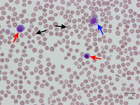
Peripheral blood smear showing leukoerythroblastic reaction: teardrop red blood cells (black arrows), myelocyte (red arrow), and promyelocyte (blue arrow)
From the collection of A. Emadi and J.L. Spivak; used with permission
See this image in context in the following section/s:
Myelofibrosis
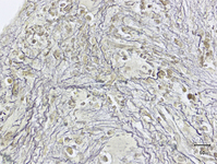
Bone marrow biopsy showing increased reticulin deposition
From the collection of A. Emadi and J.L. Spivak; used with permission
See this image in context in the following section/s:
Myelofibrosis
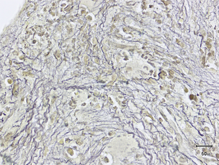
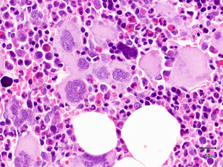
Trephine bone marrow biopsy showing megakaryocytic hyperplasia and clustering
From the collection of A. Emadi and J.L. Spivak; used with permission
See this image in context in the following section/s:
Myelofibrosis
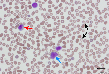
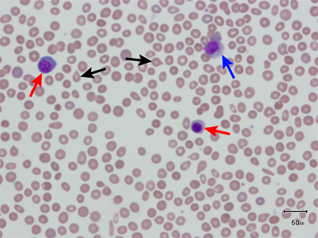
Peripheral blood smear showing teardrop red blood cells (black arrows), 2 nucleated red blood cells (red arrows), and a myelocyte (blue arrow)
From the collection of A. Emadi and J.L. Spivak; used with permission
See this image in context in the following section/s:
Use of this content is subject to our disclaimer