Etiology
Most pediatric hydroceles are congenital and, in the majority of cases, resolve within the first year of life. Most adult hydroceles are acquired. Noncommunicating hydroceles may be idiopathic or secondary to minor trauma, testicular torsion, epididymitis, varicocele operation (due to ligation of the lymphatics) or may appear as a recurrence after primary repair of a communicating or noncommunicating hydrocele.[19][20] Communicating hydroceles may occur following increased intra-abdominal fluid or pressure (due to shunts, peritoneal dialysis, or ascites) if there is a patent processus vaginalis.[6][8][21] Patients with connective tissue disorders have a high risk of communicating hydroceles.[6] Outside the US, hydroceles may also be seen in patients with filariasis as a result of lymphatic obstruction.[18]
Pathophysiology
During fetal development, an extension of the peritoneum migrates distally through the inguinal canal with the gubernaculum in the first trimester. Normally, this thin membrane that extends through the inguinal canal and descends into the scrotum (processus vaginalis) is obliterated proximally at the internal inguinal ring, and the distal portion forms the tunica vaginalis.[6][8] In the majority of cases, the processus vaginalis closes within the first year of life.[7][8][9][10] If it is not obliterated at the internal ring, it is referred to as a patent processus vaginalis, and the tunica vaginalis communicates with the peritoneum, so that peritoneal fluid flows freely between both structures and a communicating hydrocele forms. If the communication is large enough, intra-abdominal structures, such as intestine, omentum, bladder, or genital contents, may be found in the inguinal canal, and this complication is known as an indirect inguinal hernia.
While the processus vaginalis forms in both sexes in the first trimester, it does not enlarge in females. Hydrocele of the canal of Nuck is rare and results from the failure of the processus vaginalis to close, which causes fluid to accumulate within the inguinal canal.
A noncommunicating or simple hydrocele occurs in cases where the processus vaginalis is obliterated and secretion exceeds absorption of fluid from the tunica vaginalis. An abdominoscrotal hydrocele is a simple hydrocele that enlarges through the inguinal canal resulting in an abdominal component. A hydrocele of the spermatic cord is the result of segmental closure of the processus vaginalis. It is loculated and usually does not communicate with the peritoneal cavity.[22] There are two variations of a spermatic cord hydrocele; an encysted hydrocele does not communicate with the peritoneal cavity, whereas the funicular variety does communicate with the peritoneal cavity.[23][Figure caption and citation for the preceding image starts]: Normal anatomyCreated by the BMJ Group [Citation ends].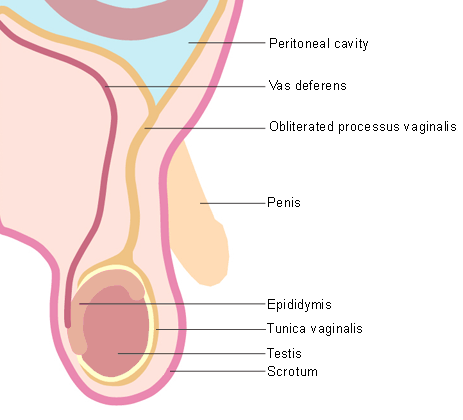 [Figure caption and citation for the preceding image starts]: Communicating hydroceleCreated by the BMJ Group [Citation ends].
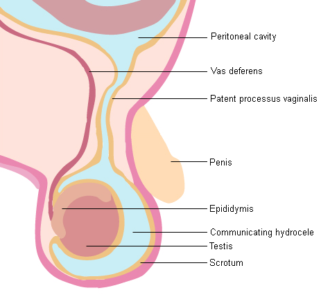 [Figure caption and citation for the preceding image starts]: Noncommunicating hydroceleCreated by the BMJ Group [Citation ends].
Classification
Common terminology
Congenital hydrocele
Results from a congenital malformation of tunica vaginalis.[1]
Acquired hydrocele[2]
Primary (or idiopathic): cause for this is unclear and is produced by defective absorption of fluid in tunica vaginalis
Secondary: caused by infection or trauma to testis.
Noncommunicating (simple) hydrocele
Accumulation of fluid around the testis without communication to the abdominal cavity.
Communicating hydrocele
Passage of peritoneal fluid to the scrotum through a patent processus vaginalis.
Abdominoscrotal hydrocele
A huge form of simple hydrocele that extends into the abdomen.
Hydrocele of the cord
A loculated hydrocele of the spermatic cord that occurs as a result of segmental closure of the processus vaginalis.[3][Figure caption and citation for the preceding image starts]: Classification of hydrocele.Created by BMJ Knowledge Centre; adapted from Tekgül S, Dogan HS, Hoebeke P, et al; European Association of Urology. Guidelines on paediatric urology. 2017 [Citation ends].

Use of this content is subject to our disclaimer