Approach
The clinical diagnostic approach to persistent pulmonary infiltrate necessitates an evaluation of host factors (age, comorbidities, immunodeficiency), the severity of symptoms, and the possibility of a noninfectious etiology. History and clinical exams, augmented by laboratory evaluation and radiographic techniques, can narrow the differential diagnosis.[14][20][21]
History
Fever and productive cough may indicate an infectious etiology, but patients with persistent pulmonary infiltrate may be asymptomatic, with only radiologic findings. Immunosuppressed patients must be evaluated initially for infection.
A history of pulmonary infection, especially with a pathogen associated with delayed radiologic resolution, can be a harbinger of slowly resolving pneumonia.
Comorbid diseases (e.g., COPD, alcohol misuse, renal failure) may also delay radiologic resolution of pneumonia.[4][6]
Epidemiologic exposure to pathogens, such as Mycobacterium tuberculosis, fungi (histoplasmosis, coccidioidomycosis, blastomycosis, and cryptococcosis), or parasites, should be evaluated.
[Figure caption and citation for the preceding image starts]: Radiographic resolution of causal pathogens of pneumoniaCreated by Athanasia Pataka [Citation ends].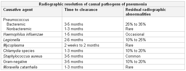 Noninfectious etiologies (e.g., radiation, drug reactions, and diffuse alveolar hemorrhage) are responsible for 20% of cases of persistent pulmonary infiltrates and must be considered Iin the absence of an infective cause.[12]
A history of myocardial infarction, angina, and peripheral edema may be pertinent in some cases of persistent pulmonary infiltrate.
Malignancies, and endobronchial obstruction from them, are a common cause of persistent pulmonary infiltrate and should always be considered in the differential diagnosis. Patients with a smoking history, hemoptysis, cachexia, or weight loss with nonresolving opacities should be evaluated for malignancy.
Hematuria may indicate alveolar hemorrhage syndromes, whereas joint pain or rashes indicate connective tissue disorders.
Asthma and persistent migratory infiltrate may suggest allergic bronchopneumonic aspergillosis.
Medication history is important, as drugs (e.g., amiodarone, bleomycin, cyclophosphamide, vincristine, taxanes) can cause pulmonary infiltrate.
Home, work environments (dust, allergens, pets) and recent travels should be assessed. Occupational and environmental exposures, radiation, and collagen-related diseases can also cause persistent pulmonary infiltrate.
Physical exam
The traditional pulmonary findings of lung disease (e.g., crackles and rales on auscultation, dullness to percussion over the chest) may be absent in the presence of persistent radiographic findings of pulmonary infiltrate. However, the examination of a patient with slowly resolving or nonresolving pneumonia should proceed in the same comprehensive manner as for any patient with a significant illness.
In immunocompetent patients with pneumonia, clinical response to antibiotic therapy is the most important determinant for further diagnostic studies for the assessment of persistent pulmonary infiltrate.[13][14] Clinical improvement and resolution of leukocytosis support response to antibiotic therapy, even when chest radiographic abnormalities persist and observation alone is reasonable.[2] When clinical improvement has not occurred and chest radiographic findings are unchanged or worse, or if there is a lack of even partial radiographic resolution by 4 weeks, further evaluation is essential, even in asymptomatic patients.[2][14]
In HIV-infected patients, skin manifestations of a pulmonary-associated bacterial, fungal, viral or neoplastic disorder may be present (e.g., cryptococcosis, Kaposi sarcoma). When a disseminated infection (fungal or mycobacterial) is suspected, an ophthalmic exam, including of the fundus and optic disc, should be performed to check for microhemorrhages.
Pulmonary congestion, peripheral edema, and elevated jugular venous pressure suggest volume overload in patients with heart failure. Clubbing of the digits may indicate idiopathic pulmonary fibrosis, asbestosis, malignancies, and (rarely) sarcoidosis or hypersensitivity pneumonitis.[72][73] Involvement of extrapulmonary sites (eyes, joints, skin, kidneys, heart, salivary glands) may suggest the presence of systemic vasculitis, sarcoidosis, and connective tissue disorders.
Laboratory
The clinical response to previous antibiotic therapy is the most important indicator of further diagnostic studies for assessing persistent pulmonary infiltrate.[13][14] In patients with nonresolving pneumonia, an initial microbiological result to confirm or deny the diagnosis of community-acquired pneumonia is essential, as the microorganism itself may explain slower resolution (e.g., Legionella pneumonia, bacteremic pneumonia). In patients with pneumonia reduction of leukocytes and C-reactive protein strongly supports response to antibiotic therapy, even with persistent chest radiographic abnormalities, and further laboratory evaluation is not necessary initially.
Microbiologic investigation may consist of:[1][13][14][68][74][75][76][77]
Sputum Gram stain
Sputum conventional bacterial culture
Direct immunofluorescence (for Legionella)
Influenza and respiratory syncytial virus (in winter months)
Sputum acid-fast bacillus smear and culture and sensitivity if tuberculosis (TB) is suspected
Nucleic acid amplification tests (should be performed on at least one respiratory specimen when a diagnosis of TB is being considered)
Stains for fungi in respiratory samples
Blood cultures
Urinary Legionella antigen (serotype 1) and Streptococcus pneumoniae antigen assays
Stains and cultures for bacteria and anaerobes in pleural fluid (if pleural effusion is present).
Multiplex polymerase chain reaction testing of lower respiratory tract samples (for viral and/or bacterial detection in patients requiring nonstandard antibiotics).
Initial and follow-up serology for Legionella species and atypical pathogens may be necessary if polymerase chain reaction testing is not available.[14] D-dimer testing can be useful to rule out pulmonary embolism.[78][79] Autoantibody testing (rheumatoid factor, antinuclear antibodies, and antineutrophil cytoplasmic autoantibodies) should be ordered if connective tissue disorders or vasculitis is suspected. Serum angiotensin-converting enzyme may be considered when sarcoidosis is entertained, although its utility is disputed.[80] Eosinophilia may suggest eosinophilic pneumonia.[81]
Imaging
Chest x-ray findings dictate the timing and choice of further evaluation for persistent pulmonary infiltrate. In most instances chest computed tomography (CT) is the most helpful radiographic modality. Infiltrations that persist without even partial resolution within 1 month or those that fail to resolve within 12 weeks should be further investigated even in asymptomatic patients.
Chest CT can detect pleural disease, complications of initial pneumonia (empyema or abscess), mediastinal abnormalities, and other noninfectious causes of persistent pulmonary infiltrate.[14] American Thoracic Society/Infectious Diseases Society of America guidelines do not recommend follow-up chest imaging in patients with community-acquired pneumonia whose symptoms are improving.[74]
Active interstitial pneumonitis may be indicated by the presence of ground-glass opacification on high-resolution chest CT, but it is not specific. CT is also useful for directing fiberoptic bronchoscopy. CT pulmonary angiography or V/Q scan is indicated if pulmonary embolism is suspected.[79]
Bronchoscopy
Fiberoptic bronchoscopy is indicated when diagnostic uncertainty remains after laboratory and radiographic evaluations are completed. Bronchoscopy has the advantage of minimal morbidity and is the preferred method of endoscopic investigation. It may be used as a diagnostic modality in approximately half of persistent pulmonary infiltrate cases.[16][82]
Bronchoscopy allows direct observation of affected areas and collection of samples. Protected brush specimen (PBS), bronchoalveolar lavage (BAL), and bronchial or transbronchial biopsy can be used. Microbiological studies of BAL and PBS may include stains and cultures for the usual bacteria, specific stains and cultures for mycobacteria, Legionella, fungi, virus; and direct immunofluorescence forLegionella. The established cut-off point to differentiate between colonization and infection is 10³ colony-forming units (cfu)/mL for PBS and 10⁴ cfu/mL for BAL fluid in immunocompetent patients, but previous antibiotic treatment should be considered. In intubated patients, endotracheal aspirates with quantitative cultures (>10⁶ cfu/mL) and sampling of distal airways (mini-BAL, protected telescoping catheter, or a blind bronchial suction sample) can be useful.
Transthoracic needle aspiration, thoracoscopic lung biopsy, or open-lung biopsy may be useful if bronchoscopy is unsuccessful or does not yield a definitive diagnosis. In patients with undiagnosed interstitial lung disease, transbronchial lung cryobiopsy has been suggested as an alternative to surgical lung biopsy if a tissue sample is required.[83]
Use of this content is subject to our disclaimer